Deck 9: B--Cardiac Physiology
ملء الشاشة (f)
سؤال
Use this figure to answer the corresponding questions.
Which structures are open during isovolumetric relaxation?


Which structures are open during isovolumetric relaxation?
سؤال
Use this figure to answer the corresponding questions.
Identify the location(s) where slow calcium channels are open.


Identify the location(s) where slow calcium channels are open.
سؤال
Use this figure to answer the corresponding questions.
Identify the location(s) where the cell's permeability to Na+ is the greatest.


Identify the location(s) where the cell's permeability to Na+ is the greatest.
سؤال
Use this figure to answer the corresponding questions.
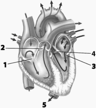
Which structures have oxygenated blood flowing through them?


Which structures have oxygenated blood flowing through them?
سؤال
Use this figure to answer the corresponding questions.
The structure labeled B

The structure labeled B
سؤال
Use this figure to answer the corresponding questions.
Identify the location(s) that indicates when fast calcium channels open within the cell.


Identify the location(s) that indicates when fast calcium channels open within the cell.
سؤال
سؤال
سؤال
Use this figure to answer the corresponding questions.
Identify the location(s) where voltage-gated potassium channels are open.

Identify the location(s) where voltage-gated potassium channels are open.
سؤال
سؤال
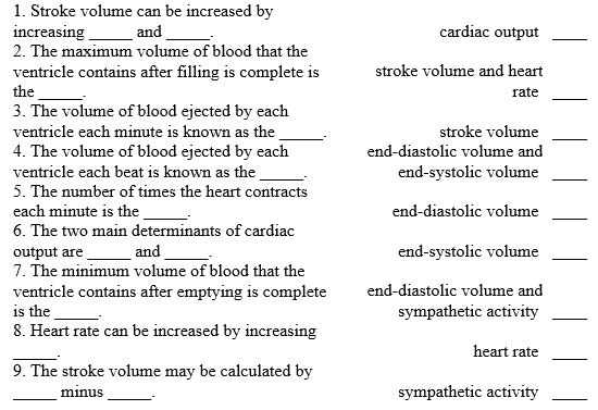
Use the answer code below to complete the following statements.
سؤال
سؤال
Use this figure to answer the corresponding questions.
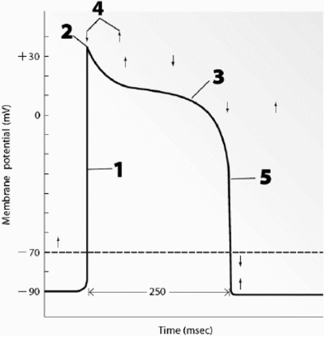
This graph shows the electrical activity for one of the heart's


This graph shows the electrical activity for one of the heart's
سؤال
Use this figure to answer the corresponding questions.
Identify the location(s) where fast calcium channels are open.
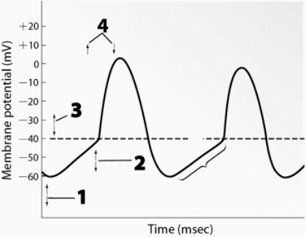

Identify the location(s) where fast calcium channels are open.
سؤال
Use this figure to answer the corresponding questions.
This graph shows the electrical activity for one of the heart's


This graph shows the electrical activity for one of the heart's
سؤال
Use this figure to answer the corresponding questions.
Identify the location(s) where fast calcium channels are open.


Identify the location(s) where fast calcium channels are open.
سؤال
Use this figure to answer the corresponding questions.
Which structures are closed during ventricular ejection?


Which structures are closed during ventricular ejection?
سؤال
سؤال
Match between columns
سؤال
Match between columns

فتح الحزمة
قم بالتسجيل لفتح البطاقات في هذه المجموعة!
Unlock Deck
Unlock Deck
1/20
العب
ملء الشاشة (f)
Deck 9: B--Cardiac Physiology
1
Use this figure to answer the corresponding questions.
Which structures are open during isovolumetric relaxation?
Which structures are open during isovolumetric relaxation?
E
2
Use this figure to answer the corresponding questions.
Identify the location(s) where slow calcium channels are open.
Identify the location(s) where slow calcium channels are open.
C
3
Use this figure to answer the corresponding questions.
Identify the location(s) where the cell's permeability to Na+ is the greatest.
Identify the location(s) where the cell's permeability to Na+ is the greatest.
A
4
Use this figure to answer the corresponding questions.
Which structures have oxygenated blood flowing through them?
Which structures have oxygenated blood flowing through them?
فتح الحزمة
افتح القفل للوصول البطاقات البالغ عددها 20 في هذه المجموعة.
فتح الحزمة
k this deck
5
Use this figure to answer the corresponding questions.
The structure labeled B
The structure labeled B
فتح الحزمة
افتح القفل للوصول البطاقات البالغ عددها 20 في هذه المجموعة.
فتح الحزمة
k this deck
6
Use this figure to answer the corresponding questions.
Identify the location(s) that indicates when fast calcium channels open within the cell.
Identify the location(s) that indicates when fast calcium channels open within the cell.
فتح الحزمة
افتح القفل للوصول البطاقات البالغ عددها 20 في هذه المجموعة.
فتح الحزمة
k this deck
7
Describe the generation of pacemaker action potentials and then track the resulting impulse through the cardiac conduction system. Include the names of specific types of channels in the first part of your answer.
فتح الحزمة
افتح القفل للوصول البطاقات البالغ عددها 20 في هذه المجموعة.
فتح الحزمة
k this deck
8
Describe the mechanisms involved by which the parasympathetic and sympathetic nervous systems affect cardiac output. Include ACh, NE, regulated K+ channels, If channels and T-type Ca2+ channels, vagus nerve, cardiac nerves, contractility, stroke volume, and heart rate in your answer.
فتح الحزمة
افتح القفل للوصول البطاقات البالغ عددها 20 في هذه المجموعة.
فتح الحزمة
k this deck
9
Use this figure to answer the corresponding questions.
Identify the location(s) where voltage-gated potassium channels are open.
Identify the location(s) where voltage-gated potassium channels are open.
فتح الحزمة
افتح القفل للوصول البطاقات البالغ عددها 20 في هذه المجموعة.
فتح الحزمة
k this deck
10
List the following in the correct order of their occurrence within the ST segment on the ECG: ventricular ejection begins, lub sound, opening of semilunar valves, period of isovolumetric contraction, closing of AV valves, ventricular systole begins.
فتح الحزمة
افتح القفل للوصول البطاقات البالغ عددها 20 في هذه المجموعة.
فتح الحزمة
k this deck
11
Use the answer code below to complete the following statements.
فتح الحزمة
افتح القفل للوصول البطاقات البالغ عددها 20 في هذه المجموعة.
فتح الحزمة
k this deck
12
Describe how contractile cells are able to contract rapidly but are not likely to experience tetanus. Include the following terms in your
فتح الحزمة
افتح القفل للوصول البطاقات البالغ عددها 20 في هذه المجموعة.
فتح الحزمة
k this deck
13
Use this figure to answer the corresponding questions.
This graph shows the electrical activity for one of the heart's
This graph shows the electrical activity for one of the heart's
فتح الحزمة
افتح القفل للوصول البطاقات البالغ عددها 20 في هذه المجموعة.
فتح الحزمة
k this deck
14
Use this figure to answer the corresponding questions.
Identify the location(s) where fast calcium channels are open.
Identify the location(s) where fast calcium channels are open.
فتح الحزمة
افتح القفل للوصول البطاقات البالغ عددها 20 في هذه المجموعة.
فتح الحزمة
k this deck
15
Use this figure to answer the corresponding questions.
This graph shows the electrical activity for one of the heart's
This graph shows the electrical activity for one of the heart's
فتح الحزمة
افتح القفل للوصول البطاقات البالغ عددها 20 في هذه المجموعة.
فتح الحزمة
k this deck
16
Use this figure to answer the corresponding questions.
Identify the location(s) where fast calcium channels are open.
Identify the location(s) where fast calcium channels are open.
فتح الحزمة
افتح القفل للوصول البطاقات البالغ عددها 20 في هذه المجموعة.
فتح الحزمة
k this deck
17
Use this figure to answer the corresponding questions.
Which structures are closed during ventricular ejection?
Which structures are closed during ventricular ejection?
فتح الحزمة
افتح القفل للوصول البطاقات البالغ عددها 20 في هذه المجموعة.
فتح الحزمة
k this deck
18
Describe the way in which high blood pressure and a defective semilunar valve can make it more difficult for the heart to pump blood into the systemic circulation and how these factors can decrease cardiac output. Include the following in your
فتح الحزمة
افتح القفل للوصول البطاقات البالغ عددها 20 في هذه المجموعة.
فتح الحزمة
k this deck
19
Match between columns
فتح الحزمة
افتح القفل للوصول البطاقات البالغ عددها 20 في هذه المجموعة.
فتح الحزمة
k this deck
20
Match between columns
فتح الحزمة
افتح القفل للوصول البطاقات البالغ عددها 20 في هذه المجموعة.
فتح الحزمة
k this deck
فتح الحزمة
افتح القفل للوصول البطاقات البالغ عددها 20 في هذه المجموعة.


