Deck 12: The Cardiovascular System: the Heart
ملء الشاشة (f)
سؤال
سؤال
سؤال
سؤال
سؤال
سؤال
سؤال
سؤال
سؤال
سؤال
سؤال
سؤال
سؤال
سؤال
سؤال
سؤال
سؤال
سؤال
سؤال
سؤال
سؤال
سؤال
سؤال
سؤال
سؤال
سؤال
سؤال
سؤال
سؤال
سؤال
سؤال
سؤال
سؤال
سؤال
سؤال
سؤال
سؤال
سؤال
سؤال
سؤال
سؤال
سؤال
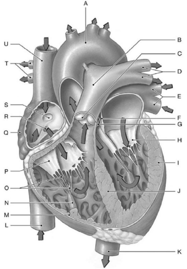 Figure 12-1 Major Internal Landmarks of the Heart
Use Figure 12-1 to identify the labeled part.
Label I represents the
A) auricle.
B) left atrium.
C) right ventricle.
D) left ventricle.
E) interventricular septum.
سؤال
Figure 12-1 Major Internal Landmarks of the HeartUse Figure 12-1 to identify the labeled part.
Label C represents the
A) tricuspid valve.
B) pulmonary (semilunar) valve.
C) aortic (semilunar) valve.
D) bicuspid valve.
E) left atrioventricular valve.
سؤال
سؤال
سؤال
سؤال
سؤال
Figure 12-1 Major Internal Landmarks of the HeartUse Figure 12-1 to identify the labeled part.
Label L represents the
A) aortic arch.
B) pulmonary trunk.
C) descending aorta.
D) superior vena cava.
E) inferior vena cava.
سؤال
Figure 12-1 Major Internal Landmarks of the HeartUse Figure 12-1 to identify the labeled part.
Label E represents the
A) pulmonary veins.
B) pulmonary trunk.
C) pulmonary arteries.
D) superior vena cava.
E) inferior vena cava.
سؤال
Figure 12-1 Major Internal Landmarks of the HeartUse Figure 12-1 to identify the labeled part.
Label D represents the
A) pulmonary veins.
B) pulmonary trunk.
C) pulmonary arteries.
D) superior vena cava.
E) inferior vena cava.
سؤال
Figure 12-1 Major Internal Landmarks of the HeartUse Figure 12-1 to identify the labeled part.
Label J represents the
A) auricle.
B) left atrium.
C) right ventricle.
D) left ventricle.
E) interventricular septum.
سؤال
سؤال
Figure 12-1 Major Internal Landmarks of the HeartUse Figure 12-1 to identify the labeled part.
Label B represents the
A) pulmonary veins.
B) pulmonary trunk.
C) pulmonary arteries.
D) superior vena cava.
E) inferior vena cava.
سؤال
Figure 12-1 Major Internal Landmarks of the HeartUse Figure 12-1 to identify the labeled part.
Label M represents the
A) auricle.
B) left atrium.
C) right ventricle.
D) left ventricle.
E) interventricular septum.
سؤال
Figure 12-1 Major Internal Landmarks of the HeartUse Figure 12-1 to identify the labeled part.
Label K represents the
A) aortic arch.
B) pulmonary trunk.
C) descending aorta.
D) superior vena cava.
E) inferior vena cava.
سؤال
Figure 12-1 Major Internal Landmarks of the HeartUse Figure 12-1 to identify the labeled part.
Label F represents the
A) interventricular septum.
B) apex.
C) opening of the coronary sinus.
D) fossa ovalis.
E) interatrial septum.
سؤال
Figure 12-1 Major Internal Landmarks of the HeartUse Figure 12-1 to identify the labeled part.
Label A represents the
A) aortic arch.
B) pulmonary trunk.
C) pulmonary arteries.
D) superior vena cava.
E) inferior vena cava.
سؤال
سؤال
Figure 12-1 Major Internal Landmarks of the HeartUse Figure 12-1 to identify the labeled part.
Label G represents the
A) tricuspid valve.
B) pulmonary (semilunar) valve.
C) aortic (semilunar) valve.
D) bicuspid valve.
E) left atrioventricular valve.
سؤال
Figure 12-1 Major Internal Landmarks of the HeartUse Figure 12-1 to identify the labeled part.
Label H represents the
A) cusps of the tricuspid valve.
B) pulmonary (semilunar) valve.
C) aortic (semilunar) valve.
D) cusps of the bicuspid valve.
E) cusps of the right atrioventricular valve.
سؤال
سؤال
Figure 12-1 Major Internal Landmarks of the HeartUse Figure 12-1 to identify the labeled part.
Label U represents the
A) right pulmonary veins.
B) pulmonary trunk.
C) right pulmonary arteries.
D) superior vena cava.
E) inferior vena cava.
سؤال
Figure 12-1 Major Internal Landmarks of the HeartUse Figure 12-1 to identify the labeled part.
Label Q represents the
A) right atrium.
B) left atrium.
C) right ventricle.
D) left ventricle.
E) interventricular septum.
سؤال
Figure 12-1 Major Internal Landmarks of the HeartUse Figure 12-1 to identify the labeled part.
Label S represents the
A) interventricular septum.
B) apex.
C) opening of the coronary sinus.
D) fossa ovalis.
E) interatrial septum.
سؤال
Figure 12-1 Major Internal Landmarks of the HeartUse Figure 12-1 to identify the labeled part.
Label R represents the
A) interventricular septum.
B) apex.
C) opening of the coronary sinus.
D) fossa ovalis.
E) interatrial septum.
سؤال
سؤال
سؤال
Figure 12-1 Major Internal Landmarks of the HeartUse Figure 12-1 to identify the labeled part.
Label T represents the
A) right pulmonary veins.
B) pulmonary trunk.
C) right pulmonary arteries.
D) superior vena cava.
E) inferior vena cava.
سؤال
سؤال
سؤال
سؤال
Figure 12-1 Major Internal Landmarks of the HeartUse Figure 12-1 to identify the labeled part.
Label N represents the
A) trabeculae carneae.
B) chordae tendineae.
C) auricle.
D) papillary muscle.
E) pectinate muscle.
سؤال
Figure 12-1 Major Internal Landmarks of the HeartUse Figure 12-1 to identify the labeled part.
Label O represents the
A) trabeculae carneae.
B) chordae tendineae.
C) auricle.
D) papillary muscle.
E) pectinate muscle.
سؤال
سؤال
سؤال
سؤال
سؤال
سؤال
سؤال
Figure 12-1 Major Internal Landmarks of the HeartUse Figure 12-1 to identify the labeled part.
Label P represents the
A) cusps of tricuspid valve.
B) pulmonary (semilunar) valve.
C) aortic (semilunar) valve.
D) cusps of bicuspid valve.
E) cusps of left atrioventricular valve.

فتح الحزمة
قم بالتسجيل لفتح البطاقات في هذه المجموعة!
Unlock Deck
Unlock Deck
1/115
العب
ملء الشاشة (f)
Deck 12: The Cardiovascular System: the Heart
1
The interventricular septum receives blood from the
A) marginal artery.
B) right coronary artery.
C) coronary sinus.
D) left coronary artery.
E) circumflex artery.
A) marginal artery.
B) right coronary artery.
C) coronary sinus.
D) left coronary artery.
E) circumflex artery.
D
2
________ permit the exchange of nutrients, dissolved gases, and waste products between the blood and surrounding tissues.
A) Veins
B) Arteries
C) Arterial trunks
D) Capillaries
E) Vena cavae
A) Veins
B) Arteries
C) Arterial trunks
D) Capillaries
E) Vena cavae
D
3
The wall between the atria is called the
A) ventricle.
B) coronary sinus.
C) coronary sulcus.
D) auricle.
E) interatrial septum.
A) ventricle.
B) coronary sinus.
C) coronary sulcus.
D) auricle.
E) interatrial septum.
E
4
The ________ circuit carries blood to and from all parts of the body except the lungs.
A) pulmonary
B) coronary
C) systemic
D) systolic
E) diastolic
A) pulmonary
B) coronary
C) systemic
D) systolic
E) diastolic
فتح الحزمة
افتح القفل للوصول البطاقات البالغ عددها 115 في هذه المجموعة.
فتح الحزمة
k this deck
5
The skeleton of the heart consists of
A) a reticular connective tissue within the myocardium.
B) a bone in the interatrial septum.
C) a bone in the interventricular septum.
D) dense bands of tough, elastic connective tissue.
E) fibrous connective tissue in the auricle of the atrium.
A) a reticular connective tissue within the myocardium.
B) a bone in the interatrial septum.
C) a bone in the interventricular septum.
D) dense bands of tough, elastic connective tissue.
E) fibrous connective tissue in the auricle of the atrium.
فتح الحزمة
افتح القفل للوصول البطاقات البالغ عددها 115 في هذه المجموعة.
فتح الحزمة
k this deck
6
Blood from the systemic circulation returns to the heart by way of the
A) coronary sinus.
B) pulmonary veins.
C) venae cavae.
D) aorta.
E) pulmonary arteries.
A) coronary sinus.
B) pulmonary veins.
C) venae cavae.
D) aorta.
E) pulmonary arteries.
فتح الحزمة
افتح القفل للوصول البطاقات البالغ عددها 115 في هذه المجموعة.
فتح الحزمة
k this deck
7
Blood flowing from the left atrium to the left ventricle flows through the ________ valve.
A) aortic
B) tricuspid
C) pulmonary
D) bicuspid
E) papillary
A) aortic
B) tricuspid
C) pulmonary
D) bicuspid
E) papillary
فتح الحزمة
افتح القفل للوصول البطاقات البالغ عددها 115 في هذه المجموعة.
فتح الحزمة
k this deck
8
The semilunar valves prevent backflow into the
A) atria.
B) aorta.
C) ventricles.
D) pulmonary trunk.
E) venae cavae.
A) atria.
B) aorta.
C) ventricles.
D) pulmonary trunk.
E) venae cavae.
فتح الحزمة
افتح القفل للوصول البطاقات البالغ عددها 115 في هذه المجموعة.
فتح الحزمة
k this deck
9
The heart is surrounded by the
A) pleural cavity.
B) peritoneal cavity.
C) cardiac skeleton.
D) pericardial cavity.
E) coronary sinus.
A) pleural cavity.
B) peritoneal cavity.
C) cardiac skeleton.
D) pericardial cavity.
E) coronary sinus.
فتح الحزمة
افتح القفل للوصول البطاقات البالغ عددها 115 في هذه المجموعة.
فتح الحزمة
k this deck
10
Each cardiac muscle cell is bound to its neighboring cells at sites called
A) intercalated discs.
B) myofibrils.
C) sarcomeres.
D) trabeculae.
E) fossa ovalis.
A) intercalated discs.
B) myofibrils.
C) sarcomeres.
D) trabeculae.
E) fossa ovalis.
فتح الحزمة
افتح القفل للوصول البطاقات البالغ عددها 115 في هذه المجموعة.
فتح الحزمة
k this deck
11
The muscle layer of the heart is the
A) epicardium.
B) myocardium.
C) endocardium.
D) visceral pericardium.
E) endothelium.
A) epicardium.
B) myocardium.
C) endocardium.
D) visceral pericardium.
E) endothelium.
فتح الحزمة
افتح القفل للوصول البطاقات البالغ عددها 115 في هذه المجموعة.
فتح الحزمة
k this deck
12
The great and middle cardiac veins drain blood directly into the
A) superior vena cava.
B) inferior vena cava.
C) coronary sinus.
D) coronary sulcus.
E) aorta.
A) superior vena cava.
B) inferior vena cava.
C) coronary sinus.
D) coronary sulcus.
E) aorta.
فتح الحزمة
افتح القفل للوصول البطاقات البالغ عددها 115 في هذه المجموعة.
فتح الحزمة
k this deck
13
The muscular ridges found on the internal surfaces of the ventricles are collectively called (the)
A) cardiac skeleton.
B) chordae tendineae.
C) papilla.
D) trabeculae carneae.
E) auricle.
A) cardiac skeleton.
B) chordae tendineae.
C) papilla.
D) trabeculae carneae.
E) auricle.
فتح الحزمة
افتح القفل للوصول البطاقات البالغ عددها 115 في هذه المجموعة.
فتح الحزمة
k this deck
14
The innermost layer of the heart wall is the
A) mediastinum.
B) parietal pericardium.
C) epicardium.
D) myocardium.
E) endocardium.
A) mediastinum.
B) parietal pericardium.
C) epicardium.
D) myocardium.
E) endocardium.
فتح الحزمة
افتح القفل للوصول البطاقات البالغ عددها 115 في هذه المجموعة.
فتح الحزمة
k this deck
15
Blood returning directly from the systemic circulation enters the
A) right atrium.
B) right ventricle.
C) left atrium.
D) left ventricle.
E) pulmonary trunk.
A) right atrium.
B) right ventricle.
C) left atrium.
D) left ventricle.
E) pulmonary trunk.
فتح الحزمة
افتح القفل للوصول البطاقات البالغ عددها 115 في هذه المجموعة.
فتح الحزمة
k this deck
16
The right atrium receives blood from the systemic circuit and pumps it to the
A) aorta.
B) left atrium.
C) right ventricle.
D) pulmonary trunk.
E) muscle tissue of the heart wall.
A) aorta.
B) left atrium.
C) right ventricle.
D) pulmonary trunk.
E) muscle tissue of the heart wall.
فتح الحزمة
افتح القفل للوصول البطاقات البالغ عددها 115 في هذه المجموعة.
فتح الحزمة
k this deck
17
The loose-fitting sac around the heart is lined by the
A) parietal pericardium.
B) epicardium.
C) endocardium.
D) parietal myocardium.
E) parietal endocardium.
A) parietal pericardium.
B) epicardium.
C) endocardium.
D) parietal myocardium.
E) parietal endocardium.
فتح الحزمة
افتح القفل للوصول البطاقات البالغ عددها 115 في هذه المجموعة.
فتح الحزمة
k this deck
18
Malfunctioning chordae tendineae would
A) allow blood to flow back into the aorta and the pulmonary trunk.
B) inhibit closure of the semilunar valves.
C) cause regurgitation into the atria.
D) enable the cusps of the AV valves to swing into the ventricles.
E) block blood flow into coronary arteries.
A) allow blood to flow back into the aorta and the pulmonary trunk.
B) inhibit closure of the semilunar valves.
C) cause regurgitation into the atria.
D) enable the cusps of the AV valves to swing into the ventricles.
E) block blood flow into coronary arteries.
فتح الحزمة
افتح القفل للوصول البطاقات البالغ عددها 115 في هذه المجموعة.
فتح الحزمة
k this deck
19
Which structural feature of the heart is a deep groove, usually filled with substantial amounts of fat, marking the border between the atria and the ventricles?
A) posterior interventricular sulcus
B) coronary sulcus
C) anterior interventricular sulcus
D) coronary sinus
E) interventricular septum
A) posterior interventricular sulcus
B) coronary sulcus
C) anterior interventricular sulcus
D) coronary sinus
E) interventricular septum
فتح الحزمة
افتح القفل للوصول البطاقات البالغ عددها 115 في هذه المجموعة.
فتح الحزمة
k this deck
20
The tricuspid valve is located
A) in the opening of the aorta.
B) in the opening of the pulmonary trunk.
C) where the vena cavae join the right atrium.
D) between the right atrium and right ventricle.
E) between the left atrium and left ventricle.
A) in the opening of the aorta.
B) in the opening of the pulmonary trunk.
C) where the vena cavae join the right atrium.
D) between the right atrium and right ventricle.
E) between the left atrium and left ventricle.
فتح الحزمة
افتح القفل للوصول البطاقات البالغ عددها 115 في هذه المجموعة.
فتح الحزمة
k this deck
21
The first blood vessels to branch from the pulmonary trunk are the
A) pulmonary arteries.
B) superior vena cavae.
C) circumflex arteries.
D) coronary arteries.
E) pulmonary veins.
A) pulmonary arteries.
B) superior vena cavae.
C) circumflex arteries.
D) coronary arteries.
E) pulmonary veins.
فتح الحزمة
افتح القفل للوصول البطاقات البالغ عددها 115 في هذه المجموعة.
فتح الحزمة
k this deck
22
The right ventricle pumps blood to the
A) left atrium.
B) left ventricle.
C) lungs.
D) systemic circuit.
E) right atrium.
A) left atrium.
B) left ventricle.
C) lungs.
D) systemic circuit.
E) right atrium.
فتح الحزمة
افتح القفل للوصول البطاقات البالغ عددها 115 في هذه المجموعة.
فتح الحزمة
k this deck
23
The function of the cardiac skeleton is to
A) convey blood away from the heart.
B) supply blood to the muscle tissue of the heart.
C) reduce friction between the opposing surfaces of the pericardial sac.
D) stabilize the position of the heart valves.
E) provide for the movement of ions and small molecules.
A) convey blood away from the heart.
B) supply blood to the muscle tissue of the heart.
C) reduce friction between the opposing surfaces of the pericardial sac.
D) stabilize the position of the heart valves.
E) provide for the movement of ions and small molecules.
فتح الحزمة
افتح القفل للوصول البطاقات البالغ عددها 115 في هذه المجموعة.
فتح الحزمة
k this deck
24
The right and left coronary arteries originate at the
A) aortic sinuses.
B) coronary sinus.
C) pulmonary trunk.
D) marginal artery.
E) fossa ovalis.
A) aortic sinuses.
B) coronary sinus.
C) pulmonary trunk.
D) marginal artery.
E) fossa ovalis.
فتح الحزمة
افتح القفل للوصول البطاقات البالغ عددها 115 في هذه المجموعة.
فتح الحزمة
k this deck
25
The atrioventricular valve on the left side of the heart is also called the
A) tricuspid valve.
B) cuspid valve.
C) mitral valve.
D) pulmonary valve.
E) aortic valve.
A) tricuspid valve.
B) cuspid valve.
C) mitral valve.
D) pulmonary valve.
E) aortic valve.
فتح الحزمة
افتح القفل للوصول البطاقات البالغ عددها 115 في هذه المجموعة.
فتح الحزمة
k this deck
26
Atrioventricular valves prevent backflow into the
A) ventricles.
B) atria.
C) venae cavae.
D) aorta.
E) pulmonary trunk.
A) ventricles.
B) atria.
C) venae cavae.
D) aorta.
E) pulmonary trunk.
فتح الحزمة
افتح القفل للوصول البطاقات البالغ عددها 115 في هذه المجموعة.
فتح الحزمة
k this deck
27
Cardiac muscle cells have abundant reserves of myoglobin, which function in
A) removing waste products.
B) storing iron.
C) removing carbon dioxide.
D) storing oxygen.
E) the shortening of individual sarcomeres.
A) removing waste products.
B) storing iron.
C) removing carbon dioxide.
D) storing oxygen.
E) the shortening of individual sarcomeres.
فتح الحزمة
افتح القفل للوصول البطاقات البالغ عددها 115 في هذه المجموعة.
فتح الحزمة
k this deck
28
Which structural feature of cardiac muscle cells enables action potentials to travel rapidly from cell to cell?
A) mitochondria
B) desmosomes
C) gap junctions
D) myofibrils
E) myoglobin
A) mitochondria
B) desmosomes
C) gap junctions
D) myofibrils
E) myoglobin
فتح الحزمة
افتح القفل للوصول البطاقات البالغ عددها 115 في هذه المجموعة.
فتح الحزمة
k this deck
29
The function of an atrium is to
A) collect blood returning to the heart.
B) pump blood to the lungs.
C) pump blood into the systemic circuit.
D) prevent the movement of blood back into the ventricles.
E) stabilize the position of the heart valves.
A) collect blood returning to the heart.
B) pump blood to the lungs.
C) pump blood into the systemic circuit.
D) prevent the movement of blood back into the ventricles.
E) stabilize the position of the heart valves.
فتح الحزمة
افتح القفل للوصول البطاقات البالغ عددها 115 في هذه المجموعة.
فتح الحزمة
k this deck
30
Which of the following correctly represents the layers associated with the heart from superficial to deep?
A) parietal pericardium, visceral pericardium, endocardium, myocardium
B) endocardium, myocardium, visceral pericardium, parietal pericardium
C) visceral pericardium, parietal pericardium, myocardium, endocardium
D) parietal pericardium, visceral pericardium, myocardium, endocardium
E) myocardium, visceral pericardium, endocardium, parietal pericardium
فتح الحزمة
افتح القفل للوصول البطاقات البالغ عددها 115 في هذه المجموعة.
فتح الحزمة
k this deck
31
Small tributaries from the branches of the coronary arteries form interconnections called
A) infarcts.
B) auricles.
C) regurgitations.
D) trabeculae carneae.
E) anastomoses.
A) infarcts.
B) auricles.
C) regurgitations.
D) trabeculae carneae.
E) anastomoses.
فتح الحزمة
افتح القفل للوصول البطاقات البالغ عددها 115 في هذه المجموعة.
فتح الحزمة
k this deck
32
The pulmonary arteries carry blood to the
A) heart.
B) lungs.
C) brain.
D) kidneys.
E) pancreas.
A) heart.
B) lungs.
C) brain.
D) kidneys.
E) pancreas.
فتح الحزمة
افتح القفل للوصول البطاقات البالغ عددها 115 في هذه المجموعة.
فتح الحزمة
k this deck
33
If valve function deteriorates such that the heart cannot maintain adequate circulatory flow, symptoms of ________ appear.
A) mitral valve prolapse
B) carditis
C) coronary artery disease
D) rheumatic fever
E) valvular heart disease
A) mitral valve prolapse
B) carditis
C) coronary artery disease
D) rheumatic fever
E) valvular heart disease
فتح الحزمة
افتح القفل للوصول البطاقات البالغ عددها 115 في هذه المجموعة.
فتح الحزمة
k this deck
34
Veins that return blood to the heart are also referred to as ________ vessels.
A) afferent
B) mitral
C) valvular
D) efferent
E) pulmonary
A) afferent
B) mitral
C) valvular
D) efferent
E) pulmonary
فتح الحزمة
افتح القفل للوصول البطاقات البالغ عددها 115 في هذه المجموعة.
فتح الحزمة
k this deck
35
The marginal artery branches off the
A) aorta.
B) left coronary artery.
C) interventricular artery.
D) coronary sinus.
E) right coronary artery.
A) aorta.
B) left coronary artery.
C) interventricular artery.
D) coronary sinus.
E) right coronary artery.
فتح الحزمة
افتح القفل للوصول البطاقات البالغ عددها 115 في هذه المجموعة.
فتح الحزمة
k this deck
36
Blood from the viscera and the lower limbs is conducted to the heart through which vessel?
A) coronary sinus
B) inferior vena cava
C) pulmonary veins
D) superior vena cava
E) cardiac vein
A) coronary sinus
B) inferior vena cava
C) pulmonary veins
D) superior vena cava
E) cardiac vein
فتح الحزمة
افتح القفل للوصول البطاقات البالغ عددها 115 في هذه المجموعة.
فتح الحزمة
k this deck
37
The semilunar valve of the left side of the heart prevents backflow from the
A) aorta.
B) pulmonary trunk.
C) pulmonary veins.
D) right ventricle.
E) left ventricle.
A) aorta.
B) pulmonary trunk.
C) pulmonary veins.
D) right ventricle.
E) left ventricle.
فتح الحزمة
افتح القفل للوصول البطاقات البالغ عددها 115 في هذه المجموعة.
فتح الحزمة
k this deck
38
The average pressure in the right ventricle is ________ the pressure in the left ventricle.
A) the same as
B) much lower than
C) slightly lower than
D) slightly higher than
E) much higher than
A) the same as
B) much lower than
C) slightly lower than
D) slightly higher than
E) much higher than
فتح الحزمة
افتح القفل للوصول البطاقات البالغ عددها 115 في هذه المجموعة.
فتح الحزمة
k this deck
39
The primary function of the venae cavae includes which of the following?
A) return blood to the right atrium
B) pump oxygenated blood into circulation
C) remove excess fluid from the heart chambers
D) anchor the heart to surrounding structures
E) prevent expansion of the heart
A) return blood to the right atrium
B) pump oxygenated blood into circulation
C) remove excess fluid from the heart chambers
D) anchor the heart to surrounding structures
E) prevent expansion of the heart
فتح الحزمة
افتح القفل للوصول البطاقات البالغ عددها 115 في هذه المجموعة.
فتح الحزمة
k this deck
40
Which of the following indicates the start of the systemic circuit?
A) pulmonary trunk
B) pulmonary arteries
C) vena cavae
D) ascending aorta
E) cardiac veins
A) pulmonary trunk
B) pulmonary arteries
C) vena cavae
D) ascending aorta
E) cardiac veins
فتح الحزمة
افتح القفل للوصول البطاقات البالغ عددها 115 في هذه المجموعة.
فتح الحزمة
k this deck
41
The left atrium receives oxygenated blood from the
A) pulmonary trunk.
B) pulmonary veins.
C) aorta.
D) inferior vena cava.
E) pulmonary arteries.
A) pulmonary trunk.
B) pulmonary veins.
C) aorta.
D) inferior vena cava.
E) pulmonary arteries.
فتح الحزمة
افتح القفل للوصول البطاقات البالغ عددها 115 في هذه المجموعة.
فتح الحزمة
k this deck
42
Figure 12-1 Major Internal Landmarks of the HeartUse Figure 12-1 to identify the labeled part.
Label I represents the
A) auricle.
B) left atrium.
C) right ventricle.
D) left ventricle.
E) interventricular septum.
فتح الحزمة
افتح القفل للوصول البطاقات البالغ عددها 115 في هذه المجموعة.
فتح الحزمة
k this deck
43
Figure 12-1 Major Internal Landmarks of the HeartUse Figure 12-1 to identify the labeled part.
Label C represents the
A) tricuspid valve.
B) pulmonary (semilunar) valve.
C) aortic (semilunar) valve.
D) bicuspid valve.
E) left atrioventricular valve.
فتح الحزمة
افتح القفل للوصول البطاقات البالغ عددها 115 في هذه المجموعة.
فتح الحزمة
k this deck
44
In ________, the cusps of the bicuspid valves do not close properly.
A) mitral valve prolapse
B) valvular heart disease
C) ventricular stenosis
D) myocardial infarctions
E) aortic sinuses
A) mitral valve prolapse
B) valvular heart disease
C) ventricular stenosis
D) myocardial infarctions
E) aortic sinuses
فتح الحزمة
افتح القفل للوصول البطاقات البالغ عددها 115 في هذه المجموعة.
فتح الحزمة
k this deck
45
A disorder resulting from the buildup of fatty deposits in the coronary arteries is called
A) valvular heart disease.
B) rheumatic heart disease.
C) coronary thrombosis.
D) coronary artery disease.
E) heart block.
A) valvular heart disease.
B) rheumatic heart disease.
C) coronary thrombosis.
D) coronary artery disease.
E) heart block.
فتح الحزمة
افتح القفل للوصول البطاقات البالغ عددها 115 في هذه المجموعة.
فتح الحزمة
k this deck
46
Deoxygenated blood is carried away from the right ventricle by the
A) pulmonary arteries.
B) pulmonary veins.
C) aorta.
D) inferior vena cava.
E) pulmonary trunk.
A) pulmonary arteries.
B) pulmonary veins.
C) aorta.
D) inferior vena cava.
E) pulmonary trunk.
فتح الحزمة
افتح القفل للوصول البطاقات البالغ عددها 115 في هذه المجموعة.
فتح الحزمة
k this deck
47
The following is a list of vessels and structures that are associated with the heart. 1. right atrium
2) left atrium
3) right ventricle
4) left ventricle
5) vena cavae
6) aorta
7) pulmonary trunk
8) pulmonary veins
What is the correct order for the flow of blood entering from the systemic circulation?
A) 1, 2, 7, 8, 3, 4, 6, 5
B) 1, 7, 3, 8, 2, 4, 6, 5
C) 5, 1, 3, 7, 8, 2, 4, 6
D) 5, 3, 1, 7, 8, 4, 2, 6
E) 5, 1, 3, 8, 7, 2, 4, 6
فتح الحزمة
افتح القفل للوصول البطاقات البالغ عددها 115 في هذه المجموعة.
فتح الحزمة
k this deck
48
Figure 12-1 Major Internal Landmarks of the HeartUse Figure 12-1 to identify the labeled part.
Label L represents the
A) aortic arch.
B) pulmonary trunk.
C) descending aorta.
D) superior vena cava.
E) inferior vena cava.
فتح الحزمة
افتح القفل للوصول البطاقات البالغ عددها 115 في هذه المجموعة.
فتح الحزمة
k this deck
49
Figure 12-1 Major Internal Landmarks of the HeartUse Figure 12-1 to identify the labeled part.
Label E represents the
A) pulmonary veins.
B) pulmonary trunk.
C) pulmonary arteries.
D) superior vena cava.
E) inferior vena cava.
فتح الحزمة
افتح القفل للوصول البطاقات البالغ عددها 115 في هذه المجموعة.
فتح الحزمة
k this deck
50
Figure 12-1 Major Internal Landmarks of the HeartUse Figure 12-1 to identify the labeled part.
Label D represents the
A) pulmonary veins.
B) pulmonary trunk.
C) pulmonary arteries.
D) superior vena cava.
E) inferior vena cava.
فتح الحزمة
افتح القفل للوصول البطاقات البالغ عددها 115 في هذه المجموعة.
فتح الحزمة
k this deck
51
Figure 12-1 Major Internal Landmarks of the HeartUse Figure 12-1 to identify the labeled part.
Label J represents the
A) auricle.
B) left atrium.
C) right ventricle.
D) left ventricle.
E) interventricular septum.
فتح الحزمة
افتح القفل للوصول البطاقات البالغ عددها 115 في هذه المجموعة.
فتح الحزمة
k this deck
52
Which statement is correct regarding the heart wall?
A) The endothelium consists of cardiac muscle tissue, blood vessels, and nerves.
B) The myocardium is comprised of a simple squamous epithelium and an underlying layer of areolar tissue.
C) The endocardium is the visceral pericardium.
D) The cardiac muscle tissue forms bands that wrap around the ventricles and spiral into the wall of the atria.
E) The epicardium is a serous membrane that consists of an exposed epithelium and an underlying layer of areolar tissue.
فتح الحزمة
افتح القفل للوصول البطاقات البالغ عددها 115 في هذه المجموعة.
فتح الحزمة
k this deck
53
Figure 12-1 Major Internal Landmarks of the HeartUse Figure 12-1 to identify the labeled part.
Label B represents the
A) pulmonary veins.
B) pulmonary trunk.
C) pulmonary arteries.
D) superior vena cava.
E) inferior vena cava.
فتح الحزمة
افتح القفل للوصول البطاقات البالغ عددها 115 في هذه المجموعة.
فتح الحزمة
k this deck
54
Figure 12-1 Major Internal Landmarks of the HeartUse Figure 12-1 to identify the labeled part.
Label M represents the
A) auricle.
B) left atrium.
C) right ventricle.
D) left ventricle.
E) interventricular septum.
فتح الحزمة
افتح القفل للوصول البطاقات البالغ عددها 115 في هذه المجموعة.
فتح الحزمة
k this deck
55
Figure 12-1 Major Internal Landmarks of the HeartUse Figure 12-1 to identify the labeled part.
Label K represents the
A) aortic arch.
B) pulmonary trunk.
C) descending aorta.
D) superior vena cava.
E) inferior vena cava.
فتح الحزمة
افتح القفل للوصول البطاقات البالغ عددها 115 في هذه المجموعة.
فتح الحزمة
k this deck
56
Figure 12-1 Major Internal Landmarks of the HeartUse Figure 12-1 to identify the labeled part.
Label F represents the
A) interventricular septum.
B) apex.
C) opening of the coronary sinus.
D) fossa ovalis.
E) interatrial septum.
فتح الحزمة
افتح القفل للوصول البطاقات البالغ عددها 115 في هذه المجموعة.
فتح الحزمة
k this deck
57
Figure 12-1 Major Internal Landmarks of the HeartUse Figure 12-1 to identify the labeled part.
Label A represents the
A) aortic arch.
B) pulmonary trunk.
C) pulmonary arteries.
D) superior vena cava.
E) inferior vena cava.
فتح الحزمة
افتح القفل للوصول البطاقات البالغ عددها 115 في هذه المجموعة.
فتح الحزمة
k this deck
58
When the ventricles are relaxed, the ________ are loose and there is no resistance to the flow of blood from atrium to ventricle.
A) auricles
B) chordae tendineae
C) papillary muscles
D) trabeculae carneae
E) aortic sinuses
A) auricles
B) chordae tendineae
C) papillary muscles
D) trabeculae carneae
E) aortic sinuses
فتح الحزمة
افتح القفل للوصول البطاقات البالغ عددها 115 في هذه المجموعة.
فتح الحزمة
k this deck
59
Figure 12-1 Major Internal Landmarks of the HeartUse Figure 12-1 to identify the labeled part.
Label G represents the
A) tricuspid valve.
B) pulmonary (semilunar) valve.
C) aortic (semilunar) valve.
D) bicuspid valve.
E) left atrioventricular valve.
فتح الحزمة
افتح القفل للوصول البطاقات البالغ عددها 115 في هذه المجموعة.
فتح الحزمة
k this deck
60
Figure 12-1 Major Internal Landmarks of the HeartUse Figure 12-1 to identify the labeled part.
Label H represents the
A) cusps of the tricuspid valve.
B) pulmonary (semilunar) valve.
C) aortic (semilunar) valve.
D) cusps of the bicuspid valve.
E) cusps of the right atrioventricular valve.
فتح الحزمة
افتح القفل للوصول البطاقات البالغ عددها 115 في هذه المجموعة.
فتح الحزمة
k this deck
61
A faster-than-normal heart rate is called
A) bradycardia.
B) tachycardia.
C) tetanus.
D) myocardial infarct.
E) arrhythmia.
A) bradycardia.
B) tachycardia.
C) tetanus.
D) myocardial infarct.
E) arrhythmia.
فتح الحزمة
افتح القفل للوصول البطاقات البالغ عددها 115 في هذه المجموعة.
فتح الحزمة
k this deck
62
Figure 12-1 Major Internal Landmarks of the HeartUse Figure 12-1 to identify the labeled part.
Label U represents the
A) right pulmonary veins.
B) pulmonary trunk.
C) right pulmonary arteries.
D) superior vena cava.
E) inferior vena cava.
فتح الحزمة
افتح القفل للوصول البطاقات البالغ عددها 115 في هذه المجموعة.
فتح الحزمة
k this deck
63
Figure 12-1 Major Internal Landmarks of the HeartUse Figure 12-1 to identify the labeled part.
Label Q represents the
A) right atrium.
B) left atrium.
C) right ventricle.
D) left ventricle.
E) interventricular septum.
فتح الحزمة
افتح القفل للوصول البطاقات البالغ عددها 115 في هذه المجموعة.
فتح الحزمة
k this deck
64
Figure 12-1 Major Internal Landmarks of the HeartUse Figure 12-1 to identify the labeled part.
Label S represents the
A) interventricular septum.
B) apex.
C) opening of the coronary sinus.
D) fossa ovalis.
E) interatrial septum.
فتح الحزمة
افتح القفل للوصول البطاقات البالغ عددها 115 في هذه المجموعة.
فتح الحزمة
k this deck
65
Figure 12-1 Major Internal Landmarks of the HeartUse Figure 12-1 to identify the labeled part.
Label R represents the
A) interventricular septum.
B) apex.
C) opening of the coronary sinus.
D) fossa ovalis.
E) interatrial septum.
فتح الحزمة
افتح القفل للوصول البطاقات البالغ عددها 115 في هذه المجموعة.
فتح الحزمة
k this deck
66
The property of heart muscle in which it contracts in the absence of neural or hormonal stimulation is called
A) tamponade.
B) thrombosis.
C) autorhythmicity.
D) arrhythmias.
E) tetanus.
A) tamponade.
B) thrombosis.
C) autorhythmicity.
D) arrhythmias.
E) tetanus.
فتح الحزمة
افتح القفل للوصول البطاقات البالغ عددها 115 في هذه المجموعة.
فتح الحزمة
k this deck
67
During rapid depolarization of a ventricular contractile cell, the voltage-gated sodium channels close when the membrane potential reaches approximately
A) -90 mV.
B) -30 mV.
C) 0 mV.
D) +30 mV.
E) +90 mV.
A) -90 mV.
B) -30 mV.
C) 0 mV.
D) +30 mV.
E) +90 mV.
فتح الحزمة
افتح القفل للوصول البطاقات البالغ عددها 115 في هذه المجموعة.
فتح الحزمة
k this deck
68
Figure 12-1 Major Internal Landmarks of the HeartUse Figure 12-1 to identify the labeled part.
Label T represents the
A) right pulmonary veins.
B) pulmonary trunk.
C) right pulmonary arteries.
D) superior vena cava.
E) inferior vena cava.
فتح الحزمة
افتح القفل للوصول البطاقات البالغ عددها 115 في هذه المجموعة.
فتح الحزمة
k this deck
69
The maximum rate of contraction in normal cardiac muscle fibers is approximately ________ per minute.
A) 80
B) 140
C) 200
D) 250
E) 300+
A) 80
B) 140
C) 200
D) 250
E) 300+
فتح الحزمة
افتح القفل للوصول البطاقات البالغ عددها 115 في هذه المجموعة.
فتح الحزمة
k this deck
70
During repolarization, ________ ions rush out of the ventricular contractile cell.
A) sodium
B) calcium
C) magnesium
D) selenium
E) potassium
A) sodium
B) calcium
C) magnesium
D) selenium
E) potassium
فتح الحزمة
افتح القفل للوصول البطاقات البالغ عددها 115 في هذه المجموعة.
فتح الحزمة
k this deck
71
The cardiac pacemaker cells are located in which of the following?
A) Purkinje fibers
B) SA node
C) AV node
D) bundle branches
E) bundle of His
A) Purkinje fibers
B) SA node
C) AV node
D) bundle branches
E) bundle of His
فتح الحزمة
افتح القفل للوصول البطاقات البالغ عددها 115 في هذه المجموعة.
فتح الحزمة
k this deck
72
Figure 12-1 Major Internal Landmarks of the HeartUse Figure 12-1 to identify the labeled part.
Label N represents the
A) trabeculae carneae.
B) chordae tendineae.
C) auricle.
D) papillary muscle.
E) pectinate muscle.
فتح الحزمة
افتح القفل للوصول البطاقات البالغ عددها 115 في هذه المجموعة.
فتح الحزمة
k this deck
73
Figure 12-1 Major Internal Landmarks of the HeartUse Figure 12-1 to identify the labeled part.
Label O represents the
A) trabeculae carneae.
B) chordae tendineae.
C) auricle.
D) papillary muscle.
E) pectinate muscle.
فتح الحزمة
افتح القفل للوصول البطاقات البالغ عددها 115 في هذه المجموعة.
فتح الحزمة
k this deck
74
Depolarization of the atria is represented on an electrocardiogram by the
A) P wave.
B) T wave.
C) S wave.
D) QRS complex.
E) PR complex.
A) P wave.
B) T wave.
C) S wave.
D) QRS complex.
E) PR complex.
فتح الحزمة
افتح القفل للوصول البطاقات البالغ عددها 115 في هذه المجموعة.
فتح الحزمة
k this deck
75
________ is a condition in which the heart rate is slower than normal.
A) Cardiac arrhythmia
B) Bradycardia
C) Tetanus
D) Summation
E) Tachycardia
A) Cardiac arrhythmia
B) Bradycardia
C) Tetanus
D) Summation
E) Tachycardia
فتح الحزمة
افتح القفل للوصول البطاقات البالغ عددها 115 في هذه المجموعة.
فتح الحزمة
k this deck
76
The QRS complex on an ECG tracing represents
A) atrial depolarization.
B) atrial repolarization.
C) ventricular depolarization.
D) ventricular repolarization.
E) ventricular contraction.
A) atrial depolarization.
B) atrial repolarization.
C) ventricular depolarization.
D) ventricular repolarization.
E) ventricular contraction.
فتح الحزمة
افتح القفل للوصول البطاقات البالغ عددها 115 في هذه المجموعة.
فتح الحزمة
k this deck
77
If the AV node does not receive action potentials generated by the SA node,
A) the ventricles will beat faster.
B) the ventricular rate of contraction will not be affected.
C) the stroke volume will increase.
D) tachycardia will occur.
E) it will establish a heart rate of 40-60 bpm.
A) the ventricles will beat faster.
B) the ventricular rate of contraction will not be affected.
C) the stroke volume will increase.
D) tachycardia will occur.
E) it will establish a heart rate of 40-60 bpm.
فتح الحزمة
افتح القفل للوصول البطاقات البالغ عددها 115 في هذه المجموعة.
فتح الحزمة
k this deck
78
Abnormal patterns of cardiac activity are known as
A) automaticity.
B) ischemias.
C) defibrillations.
D) arrhythmias.
E) contractility.
A) automaticity.
B) ischemias.
C) defibrillations.
D) arrhythmias.
E) contractility.
فتح الحزمة
افتح القفل للوصول البطاقات البالغ عددها 115 في هذه المجموعة.
فتح الحزمة
k this deck
79
The T wave of an ECG corresponds to
A) atrial depolarization.
B) atrial diastole.
C) ventricular systole.
D) ventricular repolarization.
E) atrial repolarization.
A) atrial depolarization.
B) atrial diastole.
C) ventricular systole.
D) ventricular repolarization.
E) atrial repolarization.
فتح الحزمة
افتح القفل للوصول البطاقات البالغ عددها 115 في هذه المجموعة.
فتح الحزمة
k this deck
80
Figure 12-1 Major Internal Landmarks of the HeartUse Figure 12-1 to identify the labeled part.
Label P represents the
A) cusps of tricuspid valve.
B) pulmonary (semilunar) valve.
C) aortic (semilunar) valve.
D) cusps of bicuspid valve.
E) cusps of left atrioventricular valve.
فتح الحزمة
افتح القفل للوصول البطاقات البالغ عددها 115 في هذه المجموعة.
فتح الحزمة
k this deck
فتح الحزمة
افتح القفل للوصول البطاقات البالغ عددها 115 في هذه المجموعة.


