Deck 15: Computed Tomography of the Head, Cerebral Vessels, Neck, and Spine
ملء الشاشة (f)
سؤال
سؤال
سؤال
سؤال
سؤال
سؤال
سؤال
سؤال
سؤال
سؤال
سؤال
سؤال
سؤال
سؤال
سؤال
سؤال
سؤال
سؤال
سؤال
سؤال
سؤال
The zygoma is labeled: 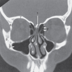
A)6.
B)7.
C)8.
D)9.
A)6.
B)7.
C)8.
D)9.
سؤال
The structure labeled 5 is the: 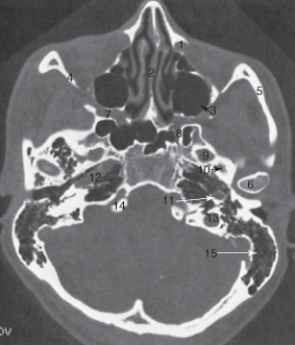
A) lateral orbital wall.
B) mandibular condyle.
C) temporal bone.
D) zygomatic arch.
A) lateral orbital wall.
B) mandibular condyle.
C) temporal bone.
D) zygomatic arch.
سؤال
The nasal septum is labeled: 
A)1.
B)2
C)3.
D)4.
A)1.
B)2
C)3.
D)4.
سؤال
The structure labeled as 1 is the: 
A) internal carotid artery.
B) masseter muscle.
C) odontoid process.
D) spinal cord.
A) internal carotid artery.
B) masseter muscle.
C) odontoid process.
D) spinal cord.
سؤال
The falx cerebri is labeled: 
A)17.
B)18.
C)19.
D)20.
A)17.
B)18.
C)19.
D)20.
سؤال
The mastoid air cells are labeled: 
A)3.
B)11.
C)12.
D)15.
A)3.
B)11.
C)12.
D)15.
سؤال
The anterior (frontal) horn of the lateral ventricle is labeled as: 
A)4.
B)8.
C)15.
D)16.
A)4.
B)8.
C)15.
D)16.
سؤال
The thalamus is labeled: 
A)9.
B)10.
C)12.
D) 14.
A)9.
B)10.
C)12.
D) 14.
سؤال
The internal jugular vein is labeled: 
A)6.
B)7.
C)8.
D)9.
A)6.
B)7.
C)8.
D)9.
سؤال
The sphenoid sinus is labeled: 
A)1.
B)2.
C)3.
D) 5.
A)1.
B)2.
C)3.
D) 5.
سؤال
The structure labeled 15 is the: 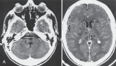
A) calcified choroid plexus.
B) fourth ventricle.
C) pons.
D) third ventricle.
A) calcified choroid plexus.
B) fourth ventricle.
C) pons.
D) third ventricle.
سؤال
The structure labeled 3 is the: 
A) cribriform plate.
B) inferior turbinate.
C) maxillary sinus.
D) middle turbinate.
A) cribriform plate.
B) inferior turbinate.
C) maxillary sinus.
D) middle turbinate.
سؤال
The structure labeled as 7 is the: 
A) cerebellar hemisphere.
B) frontal lobe.
C) middle cerebellar peduncle.
D) temporal lobe.
A) cerebellar hemisphere.
B) frontal lobe.
C) middle cerebellar peduncle.
D) temporal lobe.
سؤال
The lateral orbital wall is labeled: 
A)6.
B)7.
C)8.
D)9.
A)6.
B)7.
C)8.
D)9.
سؤال
The ethmoid sinuses are labeled: 
A)2.
B)3.
C)4.
D)5.
A)2.
B)3.
C)4.
D)5.
سؤال
The structure labeled 7 is the: 
A) cribriform plate.
B) lateral orbital wall.
C) roof of the orbit.
D) zygoma.
A) cribriform plate.
B) lateral orbital wall.
C) roof of the orbit.
D) zygoma.
سؤال
The structure labeled 3 is the: 
A) caudate nucleus.
B) fourth ventricle.
C) pons.
D) third ventricle.
A) caudate nucleus.
B) fourth ventricle.
C) pons.
D) third ventricle.
سؤال
The structure labeled as 8 is the: 
A) internal carotid artery.
B) internal jugular vein.
C) odontoid.
D) parotid gland.
A) internal carotid artery.
B) internal jugular vein.
C) odontoid.
D) parotid gland.
سؤال
The parotid gland is labeled: 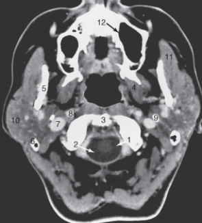
A)8.
B)9.
C)10.
D)11.
A)8.
B)9.
C)10.
D)11.
سؤال
The structure labeled 6 is the: 
A) ascending carotid canal.
B) lateral orbital wall.
C) nasal septum.
D) mandibular condyle.
A) ascending carotid canal.
B) lateral orbital wall.
C) nasal septum.
D) mandibular condyle.
سؤال
سؤال
سؤال
The intervertebral disk is labeled: 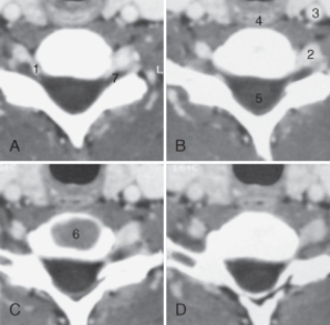
A)4
B)5.
C)6.
D)7.
A)4
B)5.
C)6.
D)7.
سؤال
The thyroid cartilage is labeled: 
A)1.
B)2.
C)3.
D)6.
A)1.
B)2.
C)3.
D)6.
سؤال
سؤال
The structure labeled as 4 is the: 
A) carotid artery.
B) esophagus.
C) spinal cord.
D) vertebral artery.
A) carotid artery.
B) esophagus.
C) spinal cord.
D) vertebral artery.
سؤال
The structure labeled as 5 is the: 
A) internal carotid artery.
B) internal jugular vein.
C) sternocleidomastoid muscle.
D) vertebral artery.
A) internal carotid artery.
B) internal jugular vein.
C) sternocleidomastoid muscle.
D) vertebral artery.
سؤال
The common carotid artery is labeled: 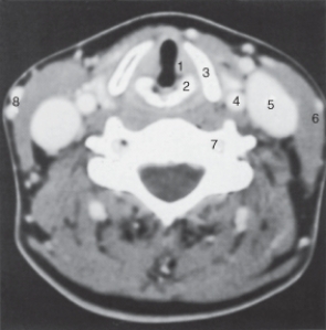
A)4.
B)5.
C)7.
D)8.
A)4.
B)5.
C)7.
D)8.
سؤال
سؤال
سؤال
سؤال
سؤال
سؤال
سؤال

فتح الحزمة
قم بالتسجيل لفتح البطاقات في هذه المجموعة!
Unlock Deck
Unlock Deck
1/55
العب
ملء الشاشة (f)
Deck 15: Computed Tomography of the Head, Cerebral Vessels, Neck, and Spine
1
Indications for a computed tomographic scan of the spine include all of the following except:
A) disk herniations.
B) osteomyelitis.
C) strokes.
D) spondylolisthesis.
A) disk herniations.
B) osteomyelitis.
C) strokes.
D) spondylolisthesis.
strokes.
2
To ensure that no life-threatening condition exists, the first examination performed should be:
A) computed tomographic angiography of the thoracic aorta.
B) computed tomographic angiography of the neck vessels.
C) computed tomographic scan of the chest.
D) computed tomographic scan of the head without contrast.
A) computed tomographic angiography of the thoracic aorta.
B) computed tomographic angiography of the neck vessels.
C) computed tomographic scan of the chest.
D) computed tomographic scan of the head without contrast.
computed tomographic scan of the head without contrast.
3
What pathologic condition describes the result of a loss of adequate blood supply to a portion of the brain?
A) Cerebral infarction
B) Hemorrhage
C) Metastatic disease
D) Tumor
A) Cerebral infarction
B) Hemorrhage
C) Metastatic disease
D) Tumor
Cerebral infarction
4
Coronal images are valuable for scans of the following except:
A) top of the cranial vault.
B) roof of the orbit.
C) skull base.
D) ventricles.
A) top of the cranial vault.
B) roof of the orbit.
C) skull base.
D) ventricles.
فتح الحزمة
افتح القفل للوصول البطاقات البالغ عددها 55 في هذه المجموعة.
فتح الحزمة
k this deck
5
Indications for a computed tomographic scan of the neck include all of the following except:
A) carotid stenosis.
B) goiter.
C) mass.
D) polyps.
A) carotid stenosis.
B) goiter.
C) mass.
D) polyps.
فتح الحزمة
افتح القفل للوصول البطاقات البالغ عددها 55 في هذه المجموعة.
فتح الحزمة
k this deck
6
Which of the following is the most common condition evaluated by repeated computed tomographic head scanning?
A) Acute subdural hematoma
B) Chronic subdural hematoma
C) Metastatic disease
D) Transient ischemic attack
A) Acute subdural hematoma
B) Chronic subdural hematoma
C) Metastatic disease
D) Transient ischemic attack
فتح الحزمة
افتح القفل للوصول البطاقات البالغ عددها 55 في هذه المجموعة.
فتح الحزمة
k this deck
7
Orbital computed tomographic images should always be available in which plane because of the pyramidal shape of the bony orbit?
A) Coronal
B) Sagittal
C) Oblique
D) Curved
A) Coronal
B) Sagittal
C) Oblique
D) Curved
فتح الحزمة
افتح القفل للوصول البطاقات البالغ عددها 55 في هذه المجموعة.
فتح الحزمة
k this deck
8
Computed tomography (CT) provides rapid information about each of the following traumatic injuries except:
A) contusions.
B) fractures.
C) hematomas.
D) metastasis.
A) contusions.
B) fractures.
C) hematomas.
D) metastasis.
فتح الحزمة
افتح القفل للوصول البطاقات البالغ عددها 55 في هذه المجموعة.
فتح الحزمة
k this deck
9
In adults, what is the preferred scanning line when imaging the head?
A) Canthomeatal line
B) Orbitomeatal line
C) Interpupillary line
D) Extrenomeatal line
A) Canthomeatal line
B) Orbitomeatal line
C) Interpupillary line
D) Extrenomeatal line
فتح الحزمة
افتح القفل للوصول البطاقات البالغ عددها 55 في هذه المجموعة.
فتح الحزمة
k this deck
10
Which images are particularly useful for assessing bone when the plane of the bone runs parallel to the axial slice?
A) Axial
B) Coronal
C) Oblique
D) Sagittal
A) Axial
B) Coronal
C) Oblique
D) Sagittal
فتح الحزمة
افتح القفل للوصول البطاقات البالغ عددها 55 في هذه المجموعة.
فتح الحزمة
k this deck
11
The matrix size on most of the current equipment is:
A) 256 * 256.
B) 512 * 512.
C) 1024*1024.
D) 2048 *2048.
A) 256 * 256.
B) 512 * 512.
C) 1024*1024.
D) 2048 *2048.
فتح الحزمة
افتح القفل للوصول البطاقات البالغ عددها 55 في هذه المجموعة.
فتح الحزمة
k this deck
12
Preparation of the patient for a computed tomographic scan of the head or spine should include all of the following except:
A) metallic objects should be removed.
B) explanation of contrast media (if indicated).
C) patient should not eat 6 hours prior to the exam.
D) patient should be made comfortable.
A) metallic objects should be removed.
B) explanation of contrast media (if indicated).
C) patient should not eat 6 hours prior to the exam.
D) patient should be made comfortable.
فتح الحزمة
افتح القفل للوصول البطاقات البالغ عددها 55 في هذه المجموعة.
فتح الحزمة
k this deck
13
Image acquisition is primarily obtained in the ___________ plane.
A) axial
B) coronal
C) oblique
D) sagittal
A) axial
B) coronal
C) oblique
D) sagittal
فتح الحزمة
افتح القفل للوصول البطاقات البالغ عددها 55 في هذه المجموعة.
فتح الحزمة
k this deck
14
All of the following are algorithms used for the brain, neck, and spine except:
A) bone.
B) detail.
C) lung.
D) standard.
A) bone.
B) detail.
C) lung.
D) standard.
فتح الحزمة
افتح القفل للوصول البطاقات البالغ عددها 55 في هذه المجموعة.
فتح الحزمة
k this deck
15
Because enhancement persists for hours, at least ___________ hours should elapse after a contrasted examination before an unenhanced scan is obtained.
A) 3-5
B) 4-6
C) 5-7
D) 6-8
A) 3-5
B) 4-6
C) 5-7
D) 6-8
فتح الحزمة
افتح القفل للوصول البطاقات البالغ عددها 55 في هذه المجموعة.
فتح الحزمة
k this deck
16
Which of the following is a form of computed tomographic angiography used in suspected acute brain infarction to determine the amount of brain tissue at risk for permanent damage?
A) Computed tomographic effusion
B) Computed tomographic venogram
C) Computed tomographic perfusion
D) Computed tomographic transmission
A) Computed tomographic effusion
B) Computed tomographic venogram
C) Computed tomographic perfusion
D) Computed tomographic transmission
فتح الحزمة
افتح القفل للوصول البطاقات البالغ عددها 55 في هذه المجموعة.
فتح الحزمة
k this deck
17
Studies requiring reformatted images are acquired with ________ slices.
A) coronal
B) sagittal
C) thick
D) thin
A) coronal
B) sagittal
C) thick
D) thin
فتح الحزمة
افتح القفل للوصول البطاقات البالغ عددها 55 في هذه المجموعة.
فتح الحزمة
k this deck
18
An important use of computed tomographic angiography of the head is in the evaluation of:
A) acute cerebral infarction.
B) fractures.
C) tumor visualization.
D) venous visualization.
A) acute cerebral infarction.
B) fractures.
C) tumor visualization.
D) venous visualization.
فتح الحزمة
افتح القفل للوصول البطاقات البالغ عددها 55 في هذه المجموعة.
فتح الحزمة
k this deck
19
The __________ plane is still the most used plane for image interpretation of the skull and its contents.
A) coronal
B) oblique
C) sagittal
D) transverse
A) coronal
B) oblique
C) sagittal
D) transverse
فتح الحزمة
افتح القفل للوصول البطاقات البالغ عددها 55 في هذه المجموعة.
فتح الحزمة
k this deck
20
The subspecialty of radiology that deals with the central nervous system (CNS) and conditions affecting the head and neck is:
A) cranial radiology.
B) gastoradiology.
C) neuroradiology.
D) vascular radiology.
A) cranial radiology.
B) gastoradiology.
C) neuroradiology.
D) vascular radiology.
فتح الحزمة
افتح القفل للوصول البطاقات البالغ عددها 55 في هذه المجموعة.
فتح الحزمة
k this deck
21
The zygoma is labeled:
A)6.
B)7.
C)8.
D)9.
A)6.
B)7.
C)8.
D)9.
فتح الحزمة
افتح القفل للوصول البطاقات البالغ عددها 55 في هذه المجموعة.
فتح الحزمة
k this deck
22
The structure labeled 5 is the:
A) lateral orbital wall.
B) mandibular condyle.
C) temporal bone.
D) zygomatic arch.
A) lateral orbital wall.
B) mandibular condyle.
C) temporal bone.
D) zygomatic arch.
فتح الحزمة
افتح القفل للوصول البطاقات البالغ عددها 55 في هذه المجموعة.
فتح الحزمة
k this deck
23
The nasal septum is labeled:
A)1.
B)2
C)3.
D)4.
A)1.
B)2
C)3.
D)4.
فتح الحزمة
افتح القفل للوصول البطاقات البالغ عددها 55 في هذه المجموعة.
فتح الحزمة
k this deck
24
The structure labeled as 1 is the:
A) internal carotid artery.
B) masseter muscle.
C) odontoid process.
D) spinal cord.
A) internal carotid artery.
B) masseter muscle.
C) odontoid process.
D) spinal cord.
فتح الحزمة
افتح القفل للوصول البطاقات البالغ عددها 55 في هذه المجموعة.
فتح الحزمة
k this deck
25
The falx cerebri is labeled:
A)17.
B)18.
C)19.
D)20.
A)17.
B)18.
C)19.
D)20.
فتح الحزمة
افتح القفل للوصول البطاقات البالغ عددها 55 في هذه المجموعة.
فتح الحزمة
k this deck
26
The mastoid air cells are labeled:
A)3.
B)11.
C)12.
D)15.
A)3.
B)11.
C)12.
D)15.
فتح الحزمة
افتح القفل للوصول البطاقات البالغ عددها 55 في هذه المجموعة.
فتح الحزمة
k this deck
27
The anterior (frontal) horn of the lateral ventricle is labeled as:
A)4.
B)8.
C)15.
D)16.
A)4.
B)8.
C)15.
D)16.
فتح الحزمة
افتح القفل للوصول البطاقات البالغ عددها 55 في هذه المجموعة.
فتح الحزمة
k this deck
28
The thalamus is labeled:
A)9.
B)10.
C)12.
D) 14.
A)9.
B)10.
C)12.
D) 14.
فتح الحزمة
افتح القفل للوصول البطاقات البالغ عددها 55 في هذه المجموعة.
فتح الحزمة
k this deck
29
The internal jugular vein is labeled:
A)6.
B)7.
C)8.
D)9.
A)6.
B)7.
C)8.
D)9.
فتح الحزمة
افتح القفل للوصول البطاقات البالغ عددها 55 في هذه المجموعة.
فتح الحزمة
k this deck
30
The sphenoid sinus is labeled:
A)1.
B)2.
C)3.
D) 5.
A)1.
B)2.
C)3.
D) 5.
فتح الحزمة
افتح القفل للوصول البطاقات البالغ عددها 55 في هذه المجموعة.
فتح الحزمة
k this deck
31
The structure labeled 15 is the:
A) calcified choroid plexus.
B) fourth ventricle.
C) pons.
D) third ventricle.
A) calcified choroid plexus.
B) fourth ventricle.
C) pons.
D) third ventricle.
فتح الحزمة
افتح القفل للوصول البطاقات البالغ عددها 55 في هذه المجموعة.
فتح الحزمة
k this deck
32
The structure labeled 3 is the:
A) cribriform plate.
B) inferior turbinate.
C) maxillary sinus.
D) middle turbinate.
A) cribriform plate.
B) inferior turbinate.
C) maxillary sinus.
D) middle turbinate.
فتح الحزمة
افتح القفل للوصول البطاقات البالغ عددها 55 في هذه المجموعة.
فتح الحزمة
k this deck
33
The structure labeled as 7 is the:
A) cerebellar hemisphere.
B) frontal lobe.
C) middle cerebellar peduncle.
D) temporal lobe.
A) cerebellar hemisphere.
B) frontal lobe.
C) middle cerebellar peduncle.
D) temporal lobe.
فتح الحزمة
افتح القفل للوصول البطاقات البالغ عددها 55 في هذه المجموعة.
فتح الحزمة
k this deck
34
The lateral orbital wall is labeled:
A)6.
B)7.
C)8.
D)9.
A)6.
B)7.
C)8.
D)9.
فتح الحزمة
افتح القفل للوصول البطاقات البالغ عددها 55 في هذه المجموعة.
فتح الحزمة
k this deck
35
The ethmoid sinuses are labeled:
A)2.
B)3.
C)4.
D)5.
A)2.
B)3.
C)4.
D)5.
فتح الحزمة
افتح القفل للوصول البطاقات البالغ عددها 55 في هذه المجموعة.
فتح الحزمة
k this deck
36
The structure labeled 7 is the:
A) cribriform plate.
B) lateral orbital wall.
C) roof of the orbit.
D) zygoma.
A) cribriform plate.
B) lateral orbital wall.
C) roof of the orbit.
D) zygoma.
فتح الحزمة
افتح القفل للوصول البطاقات البالغ عددها 55 في هذه المجموعة.
فتح الحزمة
k this deck
37
The structure labeled 3 is the:
A) caudate nucleus.
B) fourth ventricle.
C) pons.
D) third ventricle.
A) caudate nucleus.
B) fourth ventricle.
C) pons.
D) third ventricle.
فتح الحزمة
افتح القفل للوصول البطاقات البالغ عددها 55 في هذه المجموعة.
فتح الحزمة
k this deck
38
The structure labeled as 8 is the:
A) internal carotid artery.
B) internal jugular vein.
C) odontoid.
D) parotid gland.
A) internal carotid artery.
B) internal jugular vein.
C) odontoid.
D) parotid gland.
فتح الحزمة
افتح القفل للوصول البطاقات البالغ عددها 55 في هذه المجموعة.
فتح الحزمة
k this deck
39
The parotid gland is labeled:
A)8.
B)9.
C)10.
D)11.
A)8.
B)9.
C)10.
D)11.
فتح الحزمة
افتح القفل للوصول البطاقات البالغ عددها 55 في هذه المجموعة.
فتح الحزمة
k this deck
40
The structure labeled 6 is the:
A) ascending carotid canal.
B) lateral orbital wall.
C) nasal septum.
D) mandibular condyle.
A) ascending carotid canal.
B) lateral orbital wall.
C) nasal septum.
D) mandibular condyle.
فتح الحزمة
افتح القفل للوصول البطاقات البالغ عددها 55 في هذه المجموعة.
فتح الحزمة
k this deck
41
In children, a steeply angled plane is used to scan the brain to avoid radiating the ocular lens.
فتح الحزمة
افتح القفل للوصول البطاقات البالغ عددها 55 في هذه المجموعة.
فتح الحزمة
k this deck
42
Contrast enhancement is well visualized on bone algorithm or bone windows.
فتح الحزمة
افتح القفل للوصول البطاقات البالغ عددها 55 في هذه المجموعة.
فتح الحزمة
k this deck
43
The intervertebral disk is labeled:
A)4
B)5.
C)6.
D)7.
A)4
B)5.
C)6.
D)7.
فتح الحزمة
افتح القفل للوصول البطاقات البالغ عددها 55 في هذه المجموعة.
فتح الحزمة
k this deck
44
The thyroid cartilage is labeled:
A)1.
B)2.
C)3.
D)6.
A)1.
B)2.
C)3.
D)6.
فتح الحزمة
افتح القفل للوصول البطاقات البالغ عددها 55 في هذه المجموعة.
فتح الحزمة
k this deck
45
A standard dose of contrast is administered for each type of examination on the basis of the patient's weight, up to a maximum.
فتح الحزمة
افتح القفل للوصول البطاقات البالغ عددها 55 في هذه المجموعة.
فتح الحزمة
k this deck
46
The structure labeled as 4 is the:
A) carotid artery.
B) esophagus.
C) spinal cord.
D) vertebral artery.
A) carotid artery.
B) esophagus.
C) spinal cord.
D) vertebral artery.
فتح الحزمة
افتح القفل للوصول البطاقات البالغ عددها 55 في هذه المجموعة.
فتح الحزمة
k this deck
47
The structure labeled as 5 is the:
A) internal carotid artery.
B) internal jugular vein.
C) sternocleidomastoid muscle.
D) vertebral artery.
A) internal carotid artery.
B) internal jugular vein.
C) sternocleidomastoid muscle.
D) vertebral artery.
فتح الحزمة
افتح القفل للوصول البطاقات البالغ عددها 55 في هذه المجموعة.
فتح الحزمة
k this deck
48
The common carotid artery is labeled:
A)4.
B)5.
C)7.
D)8.
A)4.
B)5.
C)7.
D)8.
فتح الحزمة
افتح القفل للوصول البطاقات البالغ عددها 55 في هذه المجموعة.
فتح الحزمة
k this deck
49
CT has largely replaced plain films of the cervical spine in cases of trauma.
فتح الحزمة
افتح القفل للوصول البطاقات البالغ عددها 55 في هذه المجموعة.
فتح الحزمة
k this deck
50
Sinus disease is not well demonstrated by CT.
فتح الحزمة
افتح القفل للوصول البطاقات البالغ عددها 55 في هذه المجموعة.
فتح الحزمة
k this deck
51
Sagittal images of the temporal bone can highlight the relationships of the ossicles and the structures along the medial wall of the middle ear.
فتح الحزمة
افتح القفل للوصول البطاقات البالغ عددها 55 في هذه المجموعة.
فتح الحزمة
k this deck
52
As with plain radiographs, transverse images and coronal reformatted images are viewed as if facing the individual.
فتح الحزمة
افتح القفل للوصول البطاقات البالغ عددها 55 في هذه المجموعة.
فتح الحزمة
k this deck
53
Contrast-induced nephropathy indicates a deteriorated liver function as a result of iodine-based contrast.
فتح الحزمة
افتح القفل للوصول البطاقات البالغ عددها 55 في هذه المجموعة.
فتح الحزمة
k this deck
54
Sagittal sections are helpful in evaluating midline structures.
فتح الحزمة
افتح القفل للوصول البطاقات البالغ عددها 55 في هذه المجموعة.
فتح الحزمة
k this deck
55
A CT venogram (CTV) visualizes the jugular veins and dural sinuses for sites of possible thrombosis.
فتح الحزمة
افتح القفل للوصول البطاقات البالغ عددها 55 في هذه المجموعة.
فتح الحزمة
k this deck
فتح الحزمة
افتح القفل للوصول البطاقات البالغ عددها 55 في هذه المجموعة.


