Deck 9: Meiosis
ملء الشاشة (f)
سؤال
سؤال
سؤال
سؤال
سؤال
سؤال
سؤال
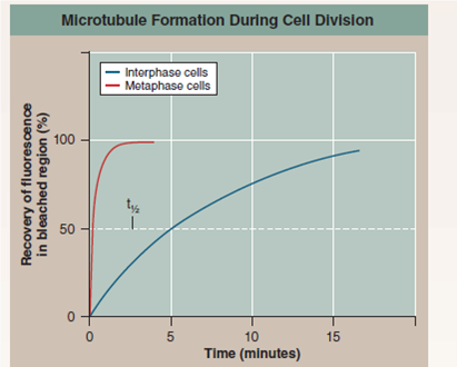
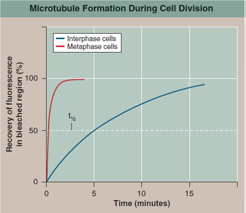
Are New Microtubules Made When the Spindle Forms?
During interphase, before the beginning of meiosis, a relatively few long microtubules extend from the centrosome (a zone around the centrioles of animal cells where microtubules are organized) to the cell periphery. Like most microtubules, these are refreshed at a low rate with resynthesis. Late in prophase, however, a dramatic change is seen-the centrosome divides into two, and a large increase is seen in the number of microtubules radiating from each of the two daughter centrosomes. The two clusters of new microtubules are easily seen as the green fibers connecting to the two sets of purple daughter chromosomes in the micrograph of early prophase below (a micrograph is a photo taken through a microscope). This burst of microtubule assembly marks the beginning of the formation of the spindle characteristic of metaphase. When it first became known to cell biologists, they asked whether these were existing microtubules being repositioned in the spindle, or newly synthesized microtubules only produced just before metaphase begins.
The graph to the upper right displays the results of an experiment designed to answer this question. Mammalian cells in culture (cells in culture are growing in the laboratory on artificial medium) were injected with microtubule subunits (tubulin) to which a fluorescent dye had been attached (a fluorescent dye is one that glows when exposed to ultraviolet or shortwavelength visual light). After the fluorescent subunits had become incorporated into the cells' microtubules, all the fluorescence in a small region of a cell was bleached by an intense laser beam, destroying the microtubules there. Any subsequent rebuilding of microtubules in the bleached region would have to employ the fluorescent subunits present in the cell, causing recovery of fluorescence in the bleached region. The graph reports this recovery as a function of time, for interphase and metaphase cells. The dotted line represents the time for 50% recovery of fluorescence (t 1/2 ) (that is, t 1/2 is the time required for half of the microtubules in the region to be resynthesized).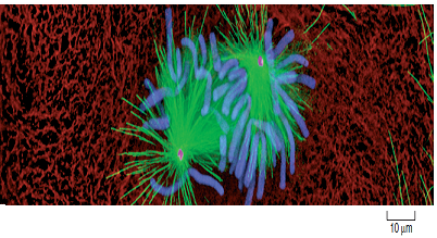

Applying Concepts
a. Variable. In the graph, what is the dependent variable?
b. t 1/2. Are new microtubules synthesized during interphase? What is the t 1/2 of this replacement synthesis? Are new microtubules synthesized during metaphase? What is the t 1/2 of this replacement synthesis?
During interphase, before the beginning of meiosis, a relatively few long microtubules extend from the centrosome (a zone around the centrioles of animal cells where microtubules are organized) to the cell periphery. Like most microtubules, these are refreshed at a low rate with resynthesis. Late in prophase, however, a dramatic change is seen-the centrosome divides into two, and a large increase is seen in the number of microtubules radiating from each of the two daughter centrosomes. The two clusters of new microtubules are easily seen as the green fibers connecting to the two sets of purple daughter chromosomes in the micrograph of early prophase below (a micrograph is a photo taken through a microscope). This burst of microtubule assembly marks the beginning of the formation of the spindle characteristic of metaphase. When it first became known to cell biologists, they asked whether these were existing microtubules being repositioned in the spindle, or newly synthesized microtubules only produced just before metaphase begins.
The graph to the upper right displays the results of an experiment designed to answer this question. Mammalian cells in culture (cells in culture are growing in the laboratory on artificial medium) were injected with microtubule subunits (tubulin) to which a fluorescent dye had been attached (a fluorescent dye is one that glows when exposed to ultraviolet or shortwavelength visual light). After the fluorescent subunits had become incorporated into the cells' microtubules, all the fluorescence in a small region of a cell was bleached by an intense laser beam, destroying the microtubules there. Any subsequent rebuilding of microtubules in the bleached region would have to employ the fluorescent subunits present in the cell, causing recovery of fluorescence in the bleached region. The graph reports this recovery as a function of time, for interphase and metaphase cells. The dotted line represents the time for 50% recovery of fluorescence (t 1/2 ) (that is, t 1/2 is the time required for half of the microtubules in the region to be resynthesized).
Applying Concepts
a. Variable. In the graph, what is the dependent variable?
b. t 1/2. Are new microtubules synthesized during interphase? What is the t 1/2 of this replacement synthesis? Are new microtubules synthesized during metaphase? What is the t 1/2 of this replacement synthesis?
سؤال
Are New Microtubules Made When the Spindle Forms?
During interphase, before the beginning of meiosis, relatively few long microtubules extend from the centrosome (a zone around the centrioles of animal cells where microtubules are organized) to the cell periphery. Like most microtubules, these are refreshed at a low rate with resynthesis. Late in prophase, however, a dramatic change is seen-the centrosome divides into two, and a large increase is seen in the number of microtubules radiating from each of the two daughter centrosomes. The two clusters of new microtubules are easily seen as the green fibers connecting to the two sets of purple chromosomes in the micrograph of early prophase below (a micrograph is a photo taken through a microscope). This burst of microtubule assembly marks the beginning of the formation of the spindle characteristic of prophase and metaphase. When it first became known to cell biologists, they asked whether these were existing microtubules being repositioned in the spindle or newly synthesized microtubules only produced just before metaphase begins.
The graph to the upper right displays the results of an experiment designed to answer this question. Mammalian cells in culture (cells in culture are growing in the laboratory on artificial medium) were injected with microtubule subunits (tubulin) to which a fluorescent dye had been attached (a fluorescent dye is one that glows when exposed to ultraviolet or short-wavelength visual light). After the fluorescent subunits had become incorporated into the cells's microtubules, all the fluorescence in a small region of a cell was bleached by an intense laser beam, destroying the microtubules there. Any subsequent rebuilding of microtubules in the bleached region would have to employ the fluorescent subunits present in the cell, causing recovery of fluorescence in the bleached region. The graph reports this recovery as a function of time for interphase and metaphase cells. The dotted line represents the time for 50% recovery of fluorescence (t 1/2 ) (that is, t 1/2 is the time required for half of the microtubules in the region to be resynthesized).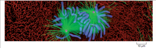

Interpreting Data Is there a difference in the rate at which microtubules are synthesized during interphase and metaphase? How big is the difference? What might account for it?
During interphase, before the beginning of meiosis, relatively few long microtubules extend from the centrosome (a zone around the centrioles of animal cells where microtubules are organized) to the cell periphery. Like most microtubules, these are refreshed at a low rate with resynthesis. Late in prophase, however, a dramatic change is seen-the centrosome divides into two, and a large increase is seen in the number of microtubules radiating from each of the two daughter centrosomes. The two clusters of new microtubules are easily seen as the green fibers connecting to the two sets of purple chromosomes in the micrograph of early prophase below (a micrograph is a photo taken through a microscope). This burst of microtubule assembly marks the beginning of the formation of the spindle characteristic of prophase and metaphase. When it first became known to cell biologists, they asked whether these were existing microtubules being repositioned in the spindle or newly synthesized microtubules only produced just before metaphase begins.
The graph to the upper right displays the results of an experiment designed to answer this question. Mammalian cells in culture (cells in culture are growing in the laboratory on artificial medium) were injected with microtubule subunits (tubulin) to which a fluorescent dye had been attached (a fluorescent dye is one that glows when exposed to ultraviolet or short-wavelength visual light). After the fluorescent subunits had become incorporated into the cells's microtubules, all the fluorescence in a small region of a cell was bleached by an intense laser beam, destroying the microtubules there. Any subsequent rebuilding of microtubules in the bleached region would have to employ the fluorescent subunits present in the cell, causing recovery of fluorescence in the bleached region. The graph reports this recovery as a function of time for interphase and metaphase cells. The dotted line represents the time for 50% recovery of fluorescence (t 1/2 ) (that is, t 1/2 is the time required for half of the microtubules in the region to be resynthesized).
Interpreting Data Is there a difference in the rate at which microtubules are synthesized during interphase and metaphase? How big is the difference? What might account for it?
سؤال
Are New Microtubules Made When the Spindle Forms?
During interphase, before the beginning of meiosis, relatively few long microtubules extend from the centrosome (a zone around the centrioles of animal cells where microtubules are organized) to the cell periphery. Like most microtubules, these are refreshed at a low rate with resynthesis. Late in prophase, however, a dramatic change is seen-the centrosome divides into two, and a large increase is seen in the number of microtubules radiating from each of the two daughter centrosomes. The two clusters of new microtubules are easily seen as the green fibers connecting to the two sets of purple chromosomes in the micrograph of early prophase below (a micrograph is a photo taken through a microscope). This burst of microtubule assembly marks the beginning of the formation of the spindle characteristic of prophase and metaphase. When it first became known to cell biologists, they asked whether these were existing microtubules being repositioned in the spindle or newly synthesized microtubules only produced just before metaphase begins.
The graph to the upper right displays the results of an experiment designed to answer this question. Mammalian cells in culture (cells in culture are growing in the laboratory on artificial medium) were injected with microtubule subunits (tubulin) to which a fluorescent dye had been attached (a fluorescent dye is one that glows when exposed to ultraviolet or short-wavelength visual light). After the fluorescent subunits had become incorporated into the cells's microtubules, all the fluorescence in a small region of a cell was bleached by an intense laser beam, destroying the microtubules there. Any subsequent rebuilding of microtubules in the bleached region would have to employ the fluorescent subunits present in the cell, causing recovery of fluorescence in the bleached region. The graph reports this recovery as a function of time for interphase and metaphase cells. The dotted line represents the time for 50% recovery of fluorescence (t 1/2 ) (that is, t 1/2 is the time required for half of the microtubules in the region to be resynthesized).
Making Inferences
a. What general statement can be made regarding the relative rates of microtubule production before and during meiosis?
b. is there any difference in the final amount of microtubule synthesis that would occur if this experiment were to be continued for an additional 15 minutes?
سؤال
Are New Microtubules Made When the Spindle Forms?
During interphase, before the beginning of meiosis, relatively few long microtubules extend from the centrosome (a zone around the centrioles of animal cells where microtubules are organized) to the cell periphery. Like most microtubules, these are refreshed at a low rate with resynthesis. Late in prophase, however, a dramatic change is seen-the centrosome divides into two, and a large increase is seen in the number of microtubules radiating from each of the two daughter centrosomes. The two clusters of new microtubules are easily seen as the green fibers connecting to the two sets of purple chromosomes in the micrograph of early prophase below (a micrograph is a photo taken through a microscope). This burst of microtubule assembly marks the beginning of the formation of the spindle characteristic of prophase and metaphase. When it first became known to cell biologists, they asked whether these were existing microtubules being repositioned in the spindle or newly synthesized microtubules only produced just before metaphase begins.
The graph to the upper right displays the results of an experiment designed to answer this question. Mammalian cells in culture (cells in culture are growing in the laboratory on artificial medium) were injected with microtubule subunits (tubulin) to which a fluorescent dye had been attached (a fluorescent dye is one that glows when exposed to ultraviolet or short-wavelength visual light). After the fluorescent subunits had become incorporated into the cells's microtubules, all the fluorescence in a small region of a cell was bleached by an intense laser beam, destroying the microtubules there. Any subsequent rebuilding of microtubules in the bleached region would have to employ the fluorescent subunits present in the cell, causing recovery of fluorescence in the bleached region. The graph reports this recovery as a function of time for interphase and metaphase cells. The dotted line represents the time for 50% recovery of fluorescence (t 1/2 ) (that is, t 1/2 is the time required for half of the microtubules in the region to be resynthesized).

Drawing Conclusions When are the microtubules of the spindle assembled?
During interphase, before the beginning of meiosis, relatively few long microtubules extend from the centrosome (a zone around the centrioles of animal cells where microtubules are organized) to the cell periphery. Like most microtubules, these are refreshed at a low rate with resynthesis. Late in prophase, however, a dramatic change is seen-the centrosome divides into two, and a large increase is seen in the number of microtubules radiating from each of the two daughter centrosomes. The two clusters of new microtubules are easily seen as the green fibers connecting to the two sets of purple chromosomes in the micrograph of early prophase below (a micrograph is a photo taken through a microscope). This burst of microtubule assembly marks the beginning of the formation of the spindle characteristic of prophase and metaphase. When it first became known to cell biologists, they asked whether these were existing microtubules being repositioned in the spindle or newly synthesized microtubules only produced just before metaphase begins.
The graph to the upper right displays the results of an experiment designed to answer this question. Mammalian cells in culture (cells in culture are growing in the laboratory on artificial medium) were injected with microtubule subunits (tubulin) to which a fluorescent dye had been attached (a fluorescent dye is one that glows when exposed to ultraviolet or short-wavelength visual light). After the fluorescent subunits had become incorporated into the cells's microtubules, all the fluorescence in a small region of a cell was bleached by an intense laser beam, destroying the microtubules there. Any subsequent rebuilding of microtubules in the bleached region would have to employ the fluorescent subunits present in the cell, causing recovery of fluorescence in the bleached region. The graph reports this recovery as a function of time for interphase and metaphase cells. The dotted line represents the time for 50% recovery of fluorescence (t 1/2 ) (that is, t 1/2 is the time required for half of the microtubules in the region to be resynthesized).
Drawing Conclusions When are the microtubules of the spindle assembled?
سؤال
Are New Microtubules Made When the Spindle Forms?
During interphase, before the beginning of meiosis, a relatively few long microtubules extend from the centrosome (a zone around the centrioles of animal cells where microtubules are organized) to the cell periphery. Like most microtubules, these are refreshed at a low rate with resynthesis. Late in prophase, however, a dramatic change is seen-the centrosome divides into two, and a large increase is seen in the number of microtubules radiating from each of the two daughter centrosomes. The two clusters of new microtubules are easily seen as the green fibers connecting to the two sets of purple daughter chromosomes in the micrograph of early prophase below (a micrograph is a photo taken through a microscope). This burst of microtubule assembly marks the beginning of the formation of the spindle characteristic of metaphase. When it first became known to cell biologists, they asked whether these were existing microtubules being repositioned in the spindle, or newly synthesized microtubules only produced just before metaphase begins.
The graph to the upper right displays the results of an experiment designed to answer this question. Mammalian cells in culture (cells in culture are growing in the laboratory on artificial medium) were injected with microtubule subunits (tubulin) to which a fluorescent dye had been attached (a fluorescent dye is one that glows when exposed to ultraviolet or shortwavelength visual light). After the fluorescent subunits had become incorporated into the cells' microtubules, all the fluorescence in a small region of a cell was bleached by an intense laser beam, destroying the microtubules there. Any subsequent rebuilding of microtubules in the bleached region would have to employ the fluorescent subunits present in the cell, causing recovery of fluorescence in the bleached region. The graph reports this recovery as a function of time, for interphase and metaphase cells. The dotted line represents the time for 50% recovery of fluorescence (t 1/2 ) (that is, t 1/2 is the time required for half of the microtubules in the region to be resynthesized).
Further Analysis The spindle breaks down after cell division is completed. Design an experiment to test whether the tubulin subunits of the spindle microtubules are recycled into other cell components, or destroyed, after meiosis.
سؤال
سؤال
سؤال
سؤال
سؤال

فتح الحزمة
قم بالتسجيل لفتح البطاقات في هذه المجموعة!
Unlock Deck
Unlock Deck
1/16
العب
ملء الشاشة (f)
Deck 9: Meiosis
1
In which stage of meiosis does crossing over occur?
A) prophase I
C) metaphase II
B) anaphase I
D) interphase
A) prophase I
C) metaphase II
B) anaphase I
D) interphase
In anaphase I one homologue with its two sister chromatids still attached moves to a pole of the cell. The other homologue moves to the opposite poles. The synthesis of DNA takes place during interphase.
In metaphase II spindle fibres bind to both the sides of the centromeres and the chromosomes gets lined up along a central plane. There is no replication of the chromosome homologues between the two nuclear divisions.
Hence the incorrect options are b, c and d.
During the process of synapsis the homologous chromosomes become closely associated along their lengths. This process of formation of complexes of homologous chromosomes is known as synapsis. The process of synapsis takes place during the prophase I of meiosis.
Hence the correct option is .
.
In metaphase II spindle fibres bind to both the sides of the centromeres and the chromosomes gets lined up along a central plane. There is no replication of the chromosome homologues between the two nuclear divisions.
Hence the incorrect options are b, c and d.
During the process of synapsis the homologous chromosomes become closely associated along their lengths. This process of formation of complexes of homologous chromosomes is known as synapsis. The process of synapsis takes place during the prophase I of meiosis.
Hence the correct option is
. 2
Mitosis results in ____, while meiosis results in _____.
A) cells genetically identical to the parent cell/haploid cells
B) haploid cells/diploid cells
C) four daughter cells/two daughter cells
D) cells with half the number of chromosomes as the parent cell/cells that vary in chromosome number
Meiosis differs from mitosis in possessing both reduction division and
A) centromere replication.
B) synapsis.
C) sister chromatids.
D) daughter cells.
In this question, we discuss the differences between mitosis and meiosis.
Meiosis occurs in two phases, both involving nuclear division. Each phase is reminiscent of mitosis in that they have a prophase, metaphase, anaphase, and telophase. DNA replication occurs in interphase (just before meiosis I), and then the process begins.
Meiosis II is almost exactly like mitosis; this phase involves the separation of sister chromatids through prophase II, metaphase II, anaphase II, and telophase II. The major difference between meiosis II and mitosis is that DNA replication does not occur - it already did before meiosis I. Homologous pairs separated during meiosis I such that each daughter cell has only one-half the number of chromosomes. Also, as a result of crossing over during prophase I, the chromosomes in the daughter cells at the end of meiosis II are not genetically identical.
a) Option A is correct - the purpose of the two processes is different, thus the different outcomes.
Option B is incorrect - mitotic division results in diploid cells.
Option C is incorrect - mitotic division results in two cells, not four.
Option D is incorrect - mitotic division results in cells with the same number of chromosomes as their parents. Improper replication results in cells with varying number of chromosomes.
b) Option B is correct - the process of synapsis is much closer in meiosis, allowing crossing over.
Option A is incorrect - centromere usage is not different in mitosis or meiosis.
Option C is incorrect - both processes have sister chromatids, they just separate at different points.
Option D is incorrect - both produce daughter cells, just different numbers.
3
Synapsis is the process whereby
A) homologous pairs of chromosomes separate and migrate toward a pole.
B) homologous chromosomes exchange chromosomal material.
C) homologous chromosomes become closely associated along their lengths.
D) the daughter cells contain half the number of chromosomes as the parent cell.
Crossing over is the process whereby
A) homologous chromosomes cross over to opposite sides of the cell.
B) homologous chromosomes exchange chromosomal material.
C) homologous chromosomes become closely associated along their lengths.
D) kinetochore fibers attach to both sides of a centromere.
In this question, we discuss the differences between mitosis and meiosis.
Meiosis occurs in two phases, both involving nuclear division. Each phase is reminiscent of mitosis in that they have a prophase, metaphase, anaphase, and telophase. DNA replication occurs in interphase (just before meiosis I), and then the process begins.
During prophase I, crossing over occurs between homologous chromosomes; sections of each homologues are physically exchanged during the crossing over process, while the homologous chromosomes are aligned with each other along their lengths. The result is recombinant chromosomes - genetic diversity. This process is the result of a difference in synpasis in meiosis; the sister chromatids are closer than in mitosis, and thus can exchange material.
a) Option C is correct - this is the definition of synapsis.
Option A is incorrect - this is part of anaphase, not synpasis.
Option B is incorrect - this is crossing over.
Option D is incorrect - this is simply the result of meiosis.
b) Option B is correct - this is the definition of crossing over.
Option A is incorrect - the crossing over is an exchange, not a change in position.
Option C is incorrect - this is the definition of synapsis.
Option D is incorrect - this is simply part of mitosis and meiosis for separation.
4
Which of the following is not a distinct feature of meiosis?
A) pairing and exchange of genetic material between homologous chromosomes
B) attachment of sister kinetochores to spindle microtubules
C) movement of sister chromatids to the same pole
D) suppression of DNA replication
In a reduction division, what gets reduced?
A) the number of chromosomes
B) the number of centromeres
C) the number of homologues
D) All of the above.
فتح الحزمة
افتح القفل للوصول البطاقات البالغ عددها 16 في هذه المجموعة.
فتح الحزمة
k this deck
5
Which of the following does not contribute to genetic diversity?
A) independent assortment
B) recombination
C) metaphase of meiosis II
D) metaphase of meiosis I
Compare independent assortment and crossing over. Which process has the greatest influence on genetic diversity?
Human beings have 23 pairs of chromosomes-22 pairs that play no role in sex determination and an XX (female) or XY (male) pair. Ignoring the effects of crossingover, what proportion of your eggs or sperm contain all of the chromosomes you received from your mother?
فتح الحزمة
افتح القفل للوصول البطاقات البالغ عددها 16 في هذه المجموعة.
فتح الحزمة
k this deck
6
A major consequence of sex and meiosis is that species
A) remain pretty much the same because the chromosomes are carefully duplicated and passed on to offspring.
B) have genetic reassortment due to processes in meiosis II.
C) have genetic reassortment due to processes in meiosis I.
D) have genetic reassortment due to processes in telophase II.
As a consequence of sex, the number of possible genetic outcomes is
A) doubled.
B) unaffected.
C) halved.
D) virtually unlimited.
فتح الحزمة
افتح القفل للوصول البطاقات البالغ عددها 16 في هذه المجموعة.
فتح الحزمة
k this deck
7
Are New Microtubules Made When the Spindle Forms?
During interphase, before the beginning of meiosis, a relatively few long microtubules extend from the centrosome (a zone around the centrioles of animal cells where microtubules are organized) to the cell periphery. Like most microtubules, these are refreshed at a low rate with resynthesis. Late in prophase, however, a dramatic change is seen-the centrosome divides into two, and a large increase is seen in the number of microtubules radiating from each of the two daughter centrosomes. The two clusters of new microtubules are easily seen as the green fibers connecting to the two sets of purple daughter chromosomes in the micrograph of early prophase below (a micrograph is a photo taken through a microscope). This burst of microtubule assembly marks the beginning of the formation of the spindle characteristic of metaphase. When it first became known to cell biologists, they asked whether these were existing microtubules being repositioned in the spindle, or newly synthesized microtubules only produced just before metaphase begins.
The graph to the upper right displays the results of an experiment designed to answer this question. Mammalian cells in culture (cells in culture are growing in the laboratory on artificial medium) were injected with microtubule subunits (tubulin) to which a fluorescent dye had been attached (a fluorescent dye is one that glows when exposed to ultraviolet or shortwavelength visual light). After the fluorescent subunits had become incorporated into the cells' microtubules, all the fluorescence in a small region of a cell was bleached by an intense laser beam, destroying the microtubules there. Any subsequent rebuilding of microtubules in the bleached region would have to employ the fluorescent subunits present in the cell, causing recovery of fluorescence in the bleached region. The graph reports this recovery as a function of time, for interphase and metaphase cells. The dotted line represents the time for 50% recovery of fluorescence (t 1/2 ) (that is, t 1/2 is the time required for half of the microtubules in the region to be resynthesized).
Applying Concepts
a. Variable. In the graph, what is the dependent variable?
b. t 1/2. Are new microtubules synthesized during interphase? What is the t 1/2 of this replacement synthesis? Are new microtubules synthesized during metaphase? What is the t 1/2 of this replacement synthesis?
فتح الحزمة
افتح القفل للوصول البطاقات البالغ عددها 16 في هذه المجموعة.
فتح الحزمة
k this deck
8
Are New Microtubules Made When the Spindle Forms?
During interphase, before the beginning of meiosis, relatively few long microtubules extend from the centrosome (a zone around the centrioles of animal cells where microtubules are organized) to the cell periphery. Like most microtubules, these are refreshed at a low rate with resynthesis. Late in prophase, however, a dramatic change is seen-the centrosome divides into two, and a large increase is seen in the number of microtubules radiating from each of the two daughter centrosomes. The two clusters of new microtubules are easily seen as the green fibers connecting to the two sets of purple chromosomes in the micrograph of early prophase below (a micrograph is a photo taken through a microscope). This burst of microtubule assembly marks the beginning of the formation of the spindle characteristic of prophase and metaphase. When it first became known to cell biologists, they asked whether these were existing microtubules being repositioned in the spindle or newly synthesized microtubules only produced just before metaphase begins.
The graph to the upper right displays the results of an experiment designed to answer this question. Mammalian cells in culture (cells in culture are growing in the laboratory on artificial medium) were injected with microtubule subunits (tubulin) to which a fluorescent dye had been attached (a fluorescent dye is one that glows when exposed to ultraviolet or short-wavelength visual light). After the fluorescent subunits had become incorporated into the cells's microtubules, all the fluorescence in a small region of a cell was bleached by an intense laser beam, destroying the microtubules there. Any subsequent rebuilding of microtubules in the bleached region would have to employ the fluorescent subunits present in the cell, causing recovery of fluorescence in the bleached region. The graph reports this recovery as a function of time for interphase and metaphase cells. The dotted line represents the time for 50% recovery of fluorescence (t 1/2 ) (that is, t 1/2 is the time required for half of the microtubules in the region to be resynthesized).
Interpreting Data Is there a difference in the rate at which microtubules are synthesized during interphase and metaphase? How big is the difference? What might account for it?
فتح الحزمة
افتح القفل للوصول البطاقات البالغ عددها 16 في هذه المجموعة.
فتح الحزمة
k this deck
9
Are New Microtubules Made When the Spindle Forms?
During interphase, before the beginning of meiosis, relatively few long microtubules extend from the centrosome (a zone around the centrioles of animal cells where microtubules are organized) to the cell periphery. Like most microtubules, these are refreshed at a low rate with resynthesis. Late in prophase, however, a dramatic change is seen-the centrosome divides into two, and a large increase is seen in the number of microtubules radiating from each of the two daughter centrosomes. The two clusters of new microtubules are easily seen as the green fibers connecting to the two sets of purple chromosomes in the micrograph of early prophase below (a micrograph is a photo taken through a microscope). This burst of microtubule assembly marks the beginning of the formation of the spindle characteristic of prophase and metaphase. When it first became known to cell biologists, they asked whether these were existing microtubules being repositioned in the spindle or newly synthesized microtubules only produced just before metaphase begins.
The graph to the upper right displays the results of an experiment designed to answer this question. Mammalian cells in culture (cells in culture are growing in the laboratory on artificial medium) were injected with microtubule subunits (tubulin) to which a fluorescent dye had been attached (a fluorescent dye is one that glows when exposed to ultraviolet or short-wavelength visual light). After the fluorescent subunits had become incorporated into the cells's microtubules, all the fluorescence in a small region of a cell was bleached by an intense laser beam, destroying the microtubules there. Any subsequent rebuilding of microtubules in the bleached region would have to employ the fluorescent subunits present in the cell, causing recovery of fluorescence in the bleached region. The graph reports this recovery as a function of time for interphase and metaphase cells. The dotted line represents the time for 50% recovery of fluorescence (t 1/2 ) (that is, t 1/2 is the time required for half of the microtubules in the region to be resynthesized).
Making Inferences
a. What general statement can be made regarding the relative rates of microtubule production before and during meiosis?
b. is there any difference in the final amount of microtubule synthesis that would occur if this experiment were to be continued for an additional 15 minutes?
فتح الحزمة
افتح القفل للوصول البطاقات البالغ عددها 16 في هذه المجموعة.
فتح الحزمة
k this deck
10
Are New Microtubules Made When the Spindle Forms?
During interphase, before the beginning of meiosis, relatively few long microtubules extend from the centrosome (a zone around the centrioles of animal cells where microtubules are organized) to the cell periphery. Like most microtubules, these are refreshed at a low rate with resynthesis. Late in prophase, however, a dramatic change is seen-the centrosome divides into two, and a large increase is seen in the number of microtubules radiating from each of the two daughter centrosomes. The two clusters of new microtubules are easily seen as the green fibers connecting to the two sets of purple chromosomes in the micrograph of early prophase below (a micrograph is a photo taken through a microscope). This burst of microtubule assembly marks the beginning of the formation of the spindle characteristic of prophase and metaphase. When it first became known to cell biologists, they asked whether these were existing microtubules being repositioned in the spindle or newly synthesized microtubules only produced just before metaphase begins.
The graph to the upper right displays the results of an experiment designed to answer this question. Mammalian cells in culture (cells in culture are growing in the laboratory on artificial medium) were injected with microtubule subunits (tubulin) to which a fluorescent dye had been attached (a fluorescent dye is one that glows when exposed to ultraviolet or short-wavelength visual light). After the fluorescent subunits had become incorporated into the cells's microtubules, all the fluorescence in a small region of a cell was bleached by an intense laser beam, destroying the microtubules there. Any subsequent rebuilding of microtubules in the bleached region would have to employ the fluorescent subunits present in the cell, causing recovery of fluorescence in the bleached region. The graph reports this recovery as a function of time for interphase and metaphase cells. The dotted line represents the time for 50% recovery of fluorescence (t 1/2 ) (that is, t 1/2 is the time required for half of the microtubules in the region to be resynthesized).
Drawing Conclusions When are the microtubules of the spindle assembled?
فتح الحزمة
افتح القفل للوصول البطاقات البالغ عددها 16 في هذه المجموعة.
فتح الحزمة
k this deck
11
Are New Microtubules Made When the Spindle Forms?
During interphase, before the beginning of meiosis, a relatively few long microtubules extend from the centrosome (a zone around the centrioles of animal cells where microtubules are organized) to the cell periphery. Like most microtubules, these are refreshed at a low rate with resynthesis. Late in prophase, however, a dramatic change is seen-the centrosome divides into two, and a large increase is seen in the number of microtubules radiating from each of the two daughter centrosomes. The two clusters of new microtubules are easily seen as the green fibers connecting to the two sets of purple daughter chromosomes in the micrograph of early prophase below (a micrograph is a photo taken through a microscope). This burst of microtubule assembly marks the beginning of the formation of the spindle characteristic of metaphase. When it first became known to cell biologists, they asked whether these were existing microtubules being repositioned in the spindle, or newly synthesized microtubules only produced just before metaphase begins.
The graph to the upper right displays the results of an experiment designed to answer this question. Mammalian cells in culture (cells in culture are growing in the laboratory on artificial medium) were injected with microtubule subunits (tubulin) to which a fluorescent dye had been attached (a fluorescent dye is one that glows when exposed to ultraviolet or shortwavelength visual light). After the fluorescent subunits had become incorporated into the cells' microtubules, all the fluorescence in a small region of a cell was bleached by an intense laser beam, destroying the microtubules there. Any subsequent rebuilding of microtubules in the bleached region would have to employ the fluorescent subunits present in the cell, causing recovery of fluorescence in the bleached region. The graph reports this recovery as a function of time, for interphase and metaphase cells. The dotted line represents the time for 50% recovery of fluorescence (t 1/2 ) (that is, t 1/2 is the time required for half of the microtubules in the region to be resynthesized).
Further Analysis The spindle breaks down after cell division is completed. Design an experiment to test whether the tubulin subunits of the spindle microtubules are recycled into other cell components, or destroyed, after meiosis.
فتح الحزمة
افتح القفل للوصول البطاقات البالغ عددها 16 في هذه المجموعة.
فتح الحزمة
k this deck
12
An egg and a sperm unite to form a new organism. To prevent the new organism from having twice as many chromosomes as its parents,
A) half of the chromosomes in the new organism quickly disassemble, leaving the correct number.
B) half of the chromosomes from the egg and half from the sperm are ejected from the new cell.
C) the large egg contains all the chromosomes, the tiny sperm only contributes some DNA.
D) the egg and sperm cells only have half the number of chromosomes found in the parents due to meiosis.
The diploid number of chromosomes in humans is 46. The haploid number is
A) 138.
B) 92.
C) 46.
D) 23.
فتح الحزمة
افتح القفل للوصول البطاقات البالغ عددها 16 في هذه المجموعة.
فتح الحزمة
k this deck
13
In organisms that have sexual life cycles, there is a time when there are
A) 1 n gametes (haploid), followed by 2 n zygotes (diploid).
B) 2 n gametes (haploid), followed by 1 n zygotes (diploid).
C) 2 n gametes (diploid), followed by 1 n zygotes (haploid).
D) 1 n gametes (diploid), followed by 2 n zygotes (haploid).
In many organisms, the haploid stage of the life cycle is dominant, with adult haploid individuals and only a brief diploid stage. No one would argue that the haploid individuals of these organisms are not alive. How then would you support or contest a statement that haploid human sperm or egg cells are not alive individuals?
فتح الحزمة
افتح القفل للوصول البطاقات البالغ عددها 16 في هذه المجموعة.
فتح الحزمة
k this deck
14
Comparing somatic cells and gametes, somatic cells are
A) diploid with one set of chromosomes.
B) haploid with one set of chromosomes.
C) diploid with two sets of chromosomes.
D) haploid with two sets of chromosomes.
A) diploid with one set of chromosomes.
B) haploid with one set of chromosomes.
C) diploid with two sets of chromosomes.
D) haploid with two sets of chromosomes.
فتح الحزمة
افتح القفل للوصول البطاقات البالغ عددها 16 في هذه المجموعة.
فتح الحزمة
k this deck
15
Which of the following occurs in meiosis I?
A) All chromosomes duplicate.
B) Homologous chromosomes randomly orient themselves on the metaphase plate, called independent assortment.
C) The duplicated sister chromatids separate.
D) The original cell divides into four diploid cells.
An organism has 56 chromosomes in its diploid stage. Indicate how many chromosomes are present in each of the following, and explain your reasoning:
A) somatic cells
B) metaphase I
C) metaphase II
D) gametes
Why is it that the sister chromatids don't separate during metaphase l as they do in mitosis?
فتح الحزمة
افتح القفل للوصول البطاقات البالغ عددها 16 في هذه المجموعة.
فتح الحزمة
k this deck
16
Which of the following occurs in meiosis II?
A) All chromosomes duplicate.
B) Homologous chromosomes randomly separate, called independent assortment.
C) The duplicated sister chromatids separate.
D) Genetically identical daughter cells are produced.
In what way is meiosis II different from mitosis?
A) Sister chromatids remain attached at the centromere.
B) Sister chromatids do not separate at anaphase II.
C) During metaphase II, spindle fibers attach to centromeres.
D) At the beginning of prophase II, sister chromatids are not genetically identical.
فتح الحزمة
افتح القفل للوصول البطاقات البالغ عددها 16 في هذه المجموعة.
فتح الحزمة
k this deck
فتح الحزمة
افتح القفل للوصول البطاقات البالغ عددها 16 في هذه المجموعة.


