Deck 6: Chest
ملء الشاشة (f)
سؤال
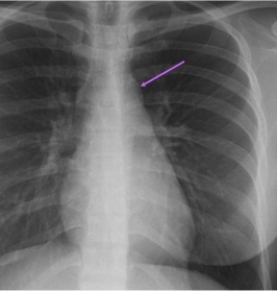 In this AP projection the arrow indicates:
A) a normal arch of aorta.
B) a dilated pulmonary trunk.
C) an enlarged tracheobronchial lymph node.
D) a trachea shifted to left due to mass effect.
سؤال
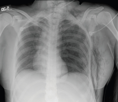
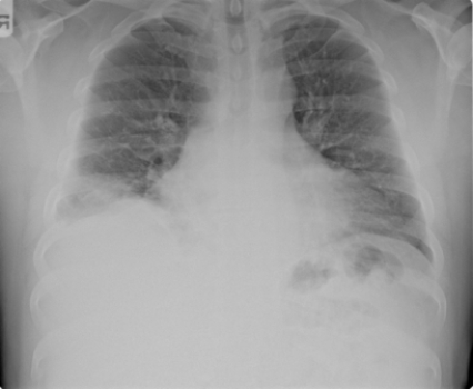
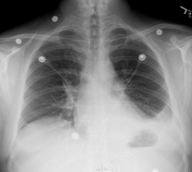 This portable AP chest radiograph shows a patient with:
A) acute pulmonary edema.
B) left lower lobe pneumonia without pleural effusion.
C) a left pleural effusion.
D) bronchogenic carcinoma in the lingula of the left upper lobe.
سؤال
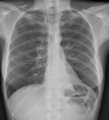
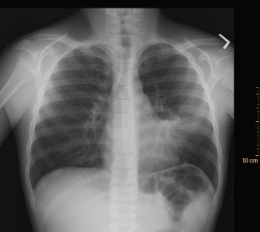 What condition does the image depict?
A) Pneumothorax
B) Pleural effusion
C) Bacterial pneumonia
D) Congestive heart failure
سؤال
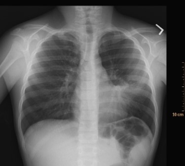 This image includes a classic radiographic finding. What is the name of this radiologic sign, and what is the most likely disease?
A) Reticulonodular pattern; interstitial lung disease
B) Silhouette sign; pneumonia
C) Cephalization of the pulmonary vascular pattern; congestive heart failure
D) Silhouette sign; metastatic malignancy
سؤال
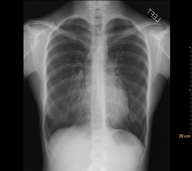 This frontal radiograph suggests an increase in lung density associated with an apparent silhouette sign along the right border of the heart. However, after a lateral view was also examined, the radiologist concluded that the patient's lungs were normal. Why?
A) The radiograph is an AP rather than the normal PA view.
B) The patient has fibrous granulomas.
C) The patient has pectus excavatum.
D) The patient is recovering from heart bypass surgery.
سؤال
 Based on the image, what does this patient likely have?
A) Left lower lung atelectasis
B) Perforated left hemidiaphragm
C) Left pleural effusion
D) Paraspinal mass
سؤال
 This patient is suffering from which symptoms?
A) Fever and cough
B) Trauma and limited air exchange on the left
C) Abdominal pain; preoperative radiograph
D) Known metastatic diseases and mediastinal adenopathy
سؤال
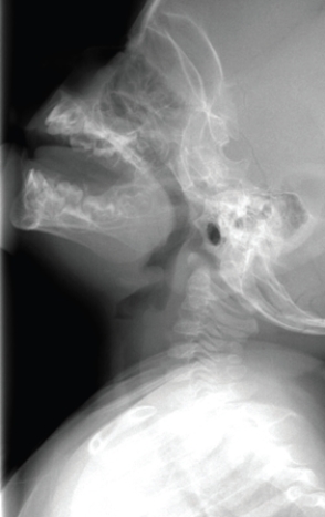 This image shows a child with:
A) allergies and enlarged adenoids causing snoring.
B) an (potentially fatal) acute epiglottitis.
C) failure to thrive and chronic dysphagia.
D) a viral illness that will respond readily to conservative treatment.
سؤال
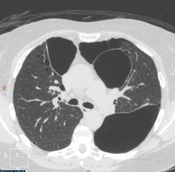 The axial CT shown depicts that this patient has which disease?
A) Emphysema
B) Interstitial lung disease
C) Nodular lung disease
D) Pneumothorax
سؤال
 Based on the image, what treatment will the patient likely receive?
A) Chemotherapy
B) Cardiac transplant
C) Surgery
D) Antibiotics
سؤال
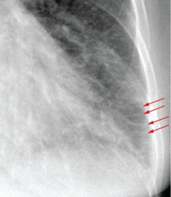 The red arrows in this image highlight a finding that is associated with:
A) congestive heart failure.
B) pneumonia.
C) silicosis.
D) emphysema.
سؤال
سؤال
سؤال
سؤال
سؤال
سؤال
سؤال
سؤال
سؤال
سؤال
سؤال
سؤال

فتح الحزمة
قم بالتسجيل لفتح البطاقات في هذه المجموعة!
Unlock Deck
Unlock Deck
1/23
العب
ملء الشاشة (f)
Deck 6: Chest
1
In this AP projection the arrow indicates:A) a normal arch of aorta.
B) a dilated pulmonary trunk.
C) an enlarged tracheobronchial lymph node.
D) a trachea shifted to left due to mass effect.
a normal arch of aorta.
2
This portable AP chest radiograph shows a patient with:A) acute pulmonary edema.
B) left lower lobe pneumonia without pleural effusion.
C) a left pleural effusion.
D) bronchogenic carcinoma in the lingula of the left upper lobe.
a left pleural effusion.
3
What condition does the image depict?A) Pneumothorax
B) Pleural effusion
C) Bacterial pneumonia
D) Congestive heart failure
Bacterial pneumonia
4
This image includes a classic radiographic finding. What is the name of this radiologic sign, and what is the most likely disease?A) Reticulonodular pattern; interstitial lung disease
B) Silhouette sign; pneumonia
C) Cephalization of the pulmonary vascular pattern; congestive heart failure
D) Silhouette sign; metastatic malignancy
فتح الحزمة
افتح القفل للوصول البطاقات البالغ عددها 23 في هذه المجموعة.
فتح الحزمة
k this deck
5
This frontal radiograph suggests an increase in lung density associated with an apparent silhouette sign along the right border of the heart. However, after a lateral view was also examined, the radiologist concluded that the patient's lungs were normal. Why?A) The radiograph is an AP rather than the normal PA view.
B) The patient has fibrous granulomas.
C) The patient has pectus excavatum.
D) The patient is recovering from heart bypass surgery.
فتح الحزمة
افتح القفل للوصول البطاقات البالغ عددها 23 في هذه المجموعة.
فتح الحزمة
k this deck
6
Based on the image, what does this patient likely have?A) Left lower lung atelectasis
B) Perforated left hemidiaphragm
C) Left pleural effusion
D) Paraspinal mass
فتح الحزمة
افتح القفل للوصول البطاقات البالغ عددها 23 في هذه المجموعة.
فتح الحزمة
k this deck
7
This patient is suffering from which symptoms?A) Fever and cough
B) Trauma and limited air exchange on the left
C) Abdominal pain; preoperative radiograph
D) Known metastatic diseases and mediastinal adenopathy
فتح الحزمة
افتح القفل للوصول البطاقات البالغ عددها 23 في هذه المجموعة.
فتح الحزمة
k this deck
8
This image shows a child with:A) allergies and enlarged adenoids causing snoring.
B) an (potentially fatal) acute epiglottitis.
C) failure to thrive and chronic dysphagia.
D) a viral illness that will respond readily to conservative treatment.
فتح الحزمة
افتح القفل للوصول البطاقات البالغ عددها 23 في هذه المجموعة.
فتح الحزمة
k this deck
9
The axial CT shown depicts that this patient has which disease?A) Emphysema
B) Interstitial lung disease
C) Nodular lung disease
D) Pneumothorax
فتح الحزمة
افتح القفل للوصول البطاقات البالغ عددها 23 في هذه المجموعة.
فتح الحزمة
k this deck
10
Based on the image, what treatment will the patient likely receive?A) Chemotherapy
B) Cardiac transplant
C) Surgery
D) Antibiotics
فتح الحزمة
افتح القفل للوصول البطاقات البالغ عددها 23 في هذه المجموعة.
فتح الحزمة
k this deck
11
The red arrows in this image highlight a finding that is associated with:A) congestive heart failure.
B) pneumonia.
C) silicosis.
D) emphysema.
فتح الحزمة
افتح القفل للوصول البطاقات البالغ عددها 23 في هذه المجموعة.
فتح الحزمة
k this deck
12
The maximum transverse diameter of the heart in a PA view should be no more than what percentage of the transverse diameter of the inner margin of the rib cage at the level of the highest point of the diaphragm to be considered normal in size?
A) 20%
B) 33%
C) 50%
D) 75%
A) 20%
B) 33%
C) 50%
D) 75%
فتح الحزمة
افتح القفل للوصول البطاقات البالغ عددها 23 في هذه المجموعة.
فتح الحزمة
k this deck
13
In examining a PA chest radiograph:
A) you should use the search pattern described by the American College of Radiology.
B) you should examine peripheral structures before more central structures.
C) you should examine superficial structures before internal structures.
D) you should use a search pattern that best suits you and one that is consistent in examining and searching for all structures.
فتح الحزمة
افتح القفل للوصول البطاقات البالغ عددها 23 في هذه المجموعة.
فتح الحزمة
k this deck
14
A blunted costophrenic (costodiaphragmatic) sulcus on a PA view is most commonly associated with:
A) pleural effusion.
B) interstitial lung disease.
C) pneumothorax.
D) emphysema.
A) pleural effusion.
B) interstitial lung disease.
C) pneumothorax.
D) emphysema.
فتح الحزمة
افتح القفل للوصول البطاقات البالغ عددها 23 في هذه المجموعة.
فتح الحزمة
k this deck
15
Which of the following statements about evaluating rib fractures is NOT true?
A) Multiple views may be required to find a nondisplaced fracture.
B) All rib fractures identified on radiographs should be followed up with CT.
C) Fractures of the first rib and an "apical cap" suggest vascular injury.
D) A flail chest refers to multiple fractures within each of three or more adjacent ribs, or three anterior ribs and sternum or costal cartilages.
فتح الحزمة
افتح القفل للوصول البطاقات البالغ عددها 23 في هذه المجموعة.
فتح الحزمة
k this deck
16
The key finding on a chest radiograph leading to the diagnosis of a pneumothorax is:
A) visibility of the visceral pleura.
B) visibility of the parietal pleura.
C) increased radiolucency of the lung.
D) decreased radiolucency of the lung.
A) visibility of the visceral pleura.
B) visibility of the parietal pleura.
C) increased radiolucency of the lung.
D) decreased radiolucency of the lung.
فتح الحزمة
افتح القفل للوصول البطاقات البالغ عددها 23 في هذه المجموعة.
فتح الحزمة
k this deck
17
Which of the following statements regarding pneumonia is CORRECT?
A) Bacterial and viral pneumonia typically are both associated with airspace opacification (consolidation).
B) Bacterial pneumonia is more typically associated with airspace opacity (consolidation) than viral pneumonia.
C) Viral pneumonia typically is associated with greater pulmonary opacity than bacterial pneumonia.
D) Neither viral nor pulmonary pneumonia have common patterns of abnormal radiographic densities.
فتح الحزمة
افتح القفل للوصول البطاقات البالغ عددها 23 في هذه المجموعة.
فتح الحزمة
k this deck
18
Croup is associated with which radiological sign?
A) Tram track
B) Air bronchogram
C) Peribronchial cuffing
D) Steeple sign
A) Tram track
B) Air bronchogram
C) Peribronchial cuffing
D) Steeple sign
فتح الحزمة
افتح القفل للوصول البطاقات البالغ عددها 23 في هذه المجموعة.
فتح الحزمة
k this deck
19
High-resolution computed tomography (HRCT) is fundamental in evaluating and diagnosing:
A) interstitial lung disease.
B) mediastinal adenopathy.
C) congestive heart failure.
D) bronchogenic carcinoma.
A) interstitial lung disease.
B) mediastinal adenopathy.
C) congestive heart failure.
D) bronchogenic carcinoma.
فتح الحزمة
افتح القفل للوصول البطاقات البالغ عددها 23 في هذه المجموعة.
فتح الحزمة
k this deck
20
Which of the following statements is NOT true for the imaging of rib metastases?
A) Radiographs will always show increased density in the rib.
B) Radiographs will always show decreased density in the rib.
C) Radionuclide bone scans will typically show foci of increased activity.
D) MRI is the procedure of choice.
A) Radiographs will always show increased density in the rib.
B) Radiographs will always show decreased density in the rib.
C) Radionuclide bone scans will typically show foci of increased activity.
D) MRI is the procedure of choice.
فتح الحزمة
افتح القفل للوصول البطاقات البالغ عددها 23 في هذه المجموعة.
فتح الحزمة
k this deck
21
Which of the following is NOT true for CCTA (cardiac or coronary CT arteriography)?
A) It is the modality of choice after a radiographic diagnosis of cardiomegaly.
B) It is less expensive than cardiac catheterization.
C) It is very sensitive for coronary artery disease.
D) It requires the IV injection of iodinated contrast material
A) It is the modality of choice after a radiographic diagnosis of cardiomegaly.
B) It is less expensive than cardiac catheterization.
C) It is very sensitive for coronary artery disease.
D) It requires the IV injection of iodinated contrast material
فتح الحزمة
افتح القفل للوصول البطاقات البالغ عددها 23 في هذه المجموعة.
فتح الحزمة
k this deck
22
The goal of a frontal chest radiograph in the setting of acute chest pain is to determine:
A) the location of a coronary infarction.
B) if there is a non-cardiac explanation for the chest pain.
C) the coronary artery dominance pattern.
D) whether the internal mammary artery can be used for coronary bypass.
A) the location of a coronary infarction.
B) if there is a non-cardiac explanation for the chest pain.
C) the coronary artery dominance pattern.
D) whether the internal mammary artery can be used for coronary bypass.
فتح الحزمة
افتح القفل للوصول البطاقات البالغ عددها 23 في هذه المجموعة.
فتح الحزمة
k this deck
23
Cephalization of the veins of the lungs is associated with all of the following EXCEPT:
A) bronchial pneumonia.
B) congestive heart failure.
C) Kerley-B lines.
D) Batwing type of pulmonary opacification.
A) bronchial pneumonia.
B) congestive heart failure.
C) Kerley-B lines.
D) Batwing type of pulmonary opacification.
فتح الحزمة
افتح القفل للوصول البطاقات البالغ عددها 23 في هذه المجموعة.
فتح الحزمة
k this deck
فتح الحزمة
افتح القفل للوصول البطاقات البالغ عددها 23 في هذه المجموعة.


