Deck 3: Spine and Spinal Cord
ملء الشاشة (f)
سؤال
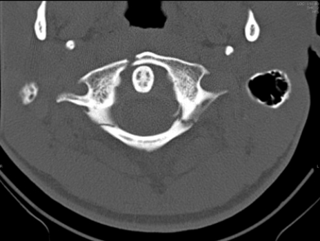 The image of this patient shows that the patient has what type of injury?
A) Hangman's fracture
B) Jefferson Burst fracture
C) Herniated C2 disk
D) C7 vertebral arch fracture
سؤال
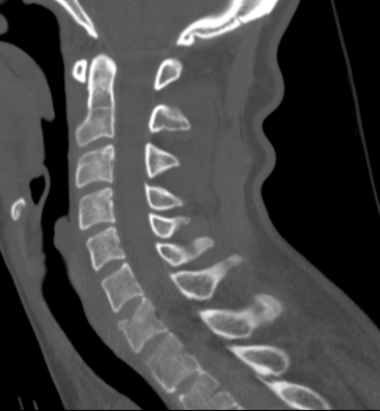 The patient in this image has which of the following?
A) A fractured arch at C1
B) A fractured hyoid bone
C) A vertebral body compression fracture
D) A spinal metastatic disease
سؤال
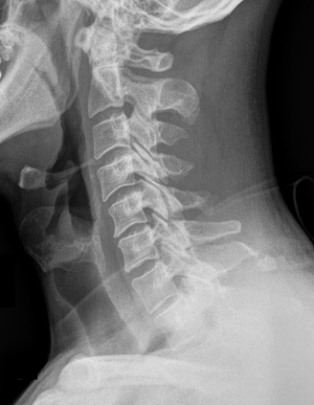 Which type of fracture(s) does the image indicate?
A) Chance fracture
B) Clay-shoveler's fracture
C) Hangman's fracture
D) Cervical facet fractures
سؤال
 This image shows that the patient has which of the following?
A) Severe dehydration of the vertebral disks
B) Transected spinal cord
C) Spinal cord tumor
D) Cerebrospinal fluid (CSF) infection
سؤال
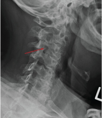 The arrow in the above image indicates:
A) a pars interarticularis defect.
B) a Chance fracture.
C) an arthritic facet joint marginal osteophyte.
D) a calcified herniated disk.
سؤال
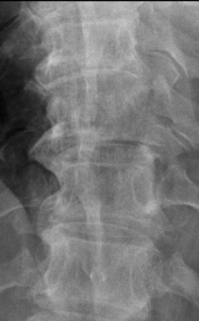 This image likely shows a patient with:
A) lytic bone disease.
B) scoliosis.
C) vertebral body compression fracture.
D) degenerative disk disease.
سؤال
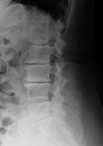 The above image depicts a patient with:
A) spondylolisthesis.
B) DISH.
C) lytic bone disease.
D) degenerative disk disease.
سؤال
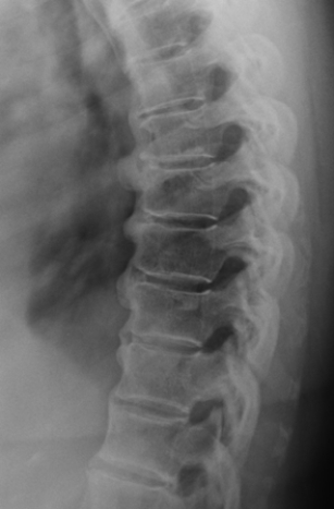 The above image depicts a patient with:
A) degenerative disk disease.
B) DISH.
C) lytic bone disease.
D) facet joint osteoarthritis.
سؤال
 Which defect is portrayed in the image of this patient?
A) An L2 pars defect
B) An L3 pars defect
C) An L4 pars defect
D) An L5 pars defect
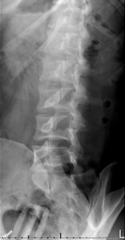
سؤال
 The image shows that this patient has:
A) anterolisthesis of the sacrum.
B) retrolisthesis of L5 in relation to the sacrum.
C) bamboo spine.
D) sacral fracture.
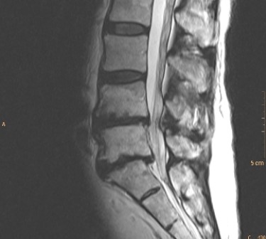
سؤال
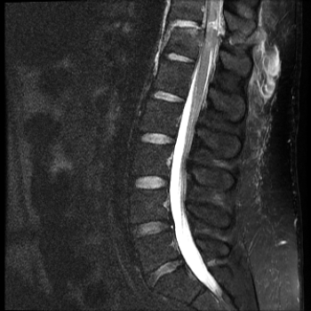
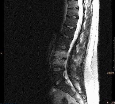 In a patient with back pain and fever, this fat suppressed T2-weighted MRI would suggest:
A) degenerative disk disease.
B) metastatic disease to the spine.
C) spondylolysis.
D) diskitis/osteomyelitis.
سؤال
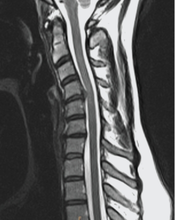 Your diagnosis based on the image shown is:
A) C4/C5 disk herniation.
B) dens fracture.
C) metastatic disease of the spine.
D) soft tissue neck injury.
سؤال
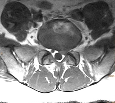 What would be your diagnosis based on the image of this patient?
A) The patient has disk herniation with nerve compression.
B) The patient has left L5/S1 facet joint osteoarthritis.
C) The patient has bilateral L5 pedicle fractures.
D) The patient has cauda equina impingement syndrome.
سؤال
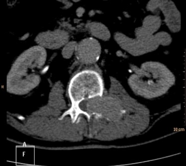 The CT shown is from a patient who presented with sciatica. Your diagnosis is most likely:
A) aortic aneurysm.
B) tumor in a pedicle.
C) herniated disk.
D) spina bifida.
سؤال
سؤال
سؤال
سؤال
سؤال
سؤال
سؤال
سؤال
سؤال
سؤال
سؤال
سؤال
سؤال
سؤال
سؤال

فتح الحزمة
قم بالتسجيل لفتح البطاقات في هذه المجموعة!
Unlock Deck
Unlock Deck
1/29
العب
ملء الشاشة (f)
Deck 3: Spine and Spinal Cord
1
The image of this patient shows that the patient has what type of injury?A) Hangman's fracture
B) Jefferson Burst fracture
C) Herniated C2 disk
D) C7 vertebral arch fracture
Jefferson Burst fracture
2
The patient in this image has which of the following?A) A fractured arch at C1
B) A fractured hyoid bone
C) A vertebral body compression fracture
D) A spinal metastatic disease
A vertebral body compression fracture
3
Which type of fracture(s) does the image indicate?A) Chance fracture
B) Clay-shoveler's fracture
C) Hangman's fracture
D) Cervical facet fractures
Clay-shoveler's fracture
4
This image shows that the patient has which of the following?A) Severe dehydration of the vertebral disks
B) Transected spinal cord
C) Spinal cord tumor
D) Cerebrospinal fluid (CSF) infection
فتح الحزمة
افتح القفل للوصول البطاقات البالغ عددها 29 في هذه المجموعة.
فتح الحزمة
k this deck
5
The arrow in the above image indicates:A) a pars interarticularis defect.
B) a Chance fracture.
C) an arthritic facet joint marginal osteophyte.
D) a calcified herniated disk.
فتح الحزمة
افتح القفل للوصول البطاقات البالغ عددها 29 في هذه المجموعة.
فتح الحزمة
k this deck
6
This image likely shows a patient with:A) lytic bone disease.
B) scoliosis.
C) vertebral body compression fracture.
D) degenerative disk disease.
فتح الحزمة
افتح القفل للوصول البطاقات البالغ عددها 29 في هذه المجموعة.
فتح الحزمة
k this deck
7
The above image depicts a patient with:A) spondylolisthesis.
B) DISH.
C) lytic bone disease.
D) degenerative disk disease.
فتح الحزمة
افتح القفل للوصول البطاقات البالغ عددها 29 في هذه المجموعة.
فتح الحزمة
k this deck
8
The above image depicts a patient with:A) degenerative disk disease.
B) DISH.
C) lytic bone disease.
D) facet joint osteoarthritis.
فتح الحزمة
افتح القفل للوصول البطاقات البالغ عددها 29 في هذه المجموعة.
فتح الحزمة
k this deck
9
Which defect is portrayed in the image of this patient?A) An L2 pars defect
B) An L3 pars defect
C) An L4 pars defect
D) An L5 pars defect
فتح الحزمة
افتح القفل للوصول البطاقات البالغ عددها 29 في هذه المجموعة.
فتح الحزمة
k this deck
10
The image shows that this patient has:A) anterolisthesis of the sacrum.
B) retrolisthesis of L5 in relation to the sacrum.
C) bamboo spine.
D) sacral fracture.
فتح الحزمة
افتح القفل للوصول البطاقات البالغ عددها 29 في هذه المجموعة.
فتح الحزمة
k this deck
11
In a patient with back pain and fever, this fat suppressed T2-weighted MRI would suggest:A) degenerative disk disease.
B) metastatic disease to the spine.
C) spondylolysis.
D) diskitis/osteomyelitis.
فتح الحزمة
افتح القفل للوصول البطاقات البالغ عددها 29 في هذه المجموعة.
فتح الحزمة
k this deck
12
Your diagnosis based on the image shown is:A) C4/C5 disk herniation.
B) dens fracture.
C) metastatic disease of the spine.
D) soft tissue neck injury.
فتح الحزمة
افتح القفل للوصول البطاقات البالغ عددها 29 في هذه المجموعة.
فتح الحزمة
k this deck
13
What would be your diagnosis based on the image of this patient?A) The patient has disk herniation with nerve compression.
B) The patient has left L5/S1 facet joint osteoarthritis.
C) The patient has bilateral L5 pedicle fractures.
D) The patient has cauda equina impingement syndrome.
فتح الحزمة
افتح القفل للوصول البطاقات البالغ عددها 29 في هذه المجموعة.
فتح الحزمة
k this deck
14
The CT shown is from a patient who presented with sciatica. Your diagnosis is most likely:A) aortic aneurysm.
B) tumor in a pedicle.
C) herniated disk.
D) spina bifida.
فتح الحزمة
افتح القفل للوصول البطاقات البالغ عددها 29 في هذه المجموعة.
فتح الحزمة
k this deck
15
Which of the following is NOT true of degenerative disk disease?
A) It is typically associated with marginal osteophytes.
B) It is typically associated with disk dehydration.
C) It is always associated with back pain.
D) It does not require advanced imaging, such as MRI, for diagnosis.
A) It is typically associated with marginal osteophytes.
B) It is typically associated with disk dehydration.
C) It is always associated with back pain.
D) It does not require advanced imaging, such as MRI, for diagnosis.
فتح الحزمة
افتح القفل للوصول البطاقات البالغ عددها 29 في هذه المجموعة.
فتح الحزمة
k this deck
16
Which of the following radiographic views BEST displays a pars interarticularis fracture?
A) AP
B) PA
C) Lateral
D) Oblique
A) AP
B) PA
C) Lateral
D) Oblique
فتح الحزمة
افتح القفل للوصول البطاقات البالغ عددها 29 في هذه المجموعة.
فتح الحزمة
k this deck
17
In patients who have severe spine trauma but do not show neurologic signs, the procedure of choice is:
A) CT.
B) MRI.
C) radiography.
D) ultrasound.
A) CT.
B) MRI.
C) radiography.
D) ultrasound.
فتح الحزمة
افتح القفل للوصول البطاقات البالغ عددها 29 في هذه المجموعة.
فتح الحزمة
k this deck
18
Which of the following cervical fractures are typically stable?
A) Teardrop
B) Bilateral facet
C) Anterior wedge compression
D) Hangman's
A) Teardrop
B) Bilateral facet
C) Anterior wedge compression
D) Hangman's
فتح الحزمة
افتح القفل للوصول البطاقات البالغ عددها 29 في هذه المجموعة.
فتح الحزمة
k this deck
19
In a patient with a Chance fracture, you would also likely suspect injury to the:
A) ribs.
B) lungs.
C) pancreas.
D) liver.
A) ribs.
B) lungs.
C) pancreas.
D) liver.
فتح الحزمة
افتح القفل للوصول البطاقات البالغ عددها 29 في هذه المجموعة.
فتح الحزمة
k this deck
20
In a patient with low back pain with or without lumbar radiculopathy, standing radiographs may be helpful because they:
A) may provide functional information regarding the alignment of vertebrae when weight bearing.
B) provide better bone detail.
C) enable more vertebrae to be seen.
D) require less ionizing radiation to produce a comparable image.
A) may provide functional information regarding the alignment of vertebrae when weight bearing.
B) provide better bone detail.
C) enable more vertebrae to be seen.
D) require less ionizing radiation to produce a comparable image.
فتح الحزمة
افتح القفل للوصول البطاقات البالغ عددها 29 في هذه المجموعة.
فتح الحزمة
k this deck
21
Which radiographic view is BEST for evaluating suspected rotational subluxation at C1/C2?
A) Open-mouth odontoid
B) Lateral C-spine with flexion and extension
C) AP view with cephalad angulation
D) Posterior oblique views
A) Open-mouth odontoid
B) Lateral C-spine with flexion and extension
C) AP view with cephalad angulation
D) Posterior oblique views
فتح الحزمة
افتح القفل للوصول البطاقات البالغ عددها 29 في هذه المجموعة.
فتح الحزمة
k this deck
22
Which of the following is NOT associated with an osteoarthritic facet joint?
A) Decreased density of subchondral bone
B) Osteophytes
C) Reduction in joint space
D) Narrowing of the neuroforamen
A) Decreased density of subchondral bone
B) Osteophytes
C) Reduction in joint space
D) Narrowing of the neuroforamen
فتح الحزمة
افتح القفل للوصول البطاقات البالغ عددها 29 في هذه المجموعة.
فتح الحزمة
k this deck
23
The pedicle sign refers to which of the following?
A) An increase in the visibility of a pedicle due to blastic disease
B) A decrease in the visibility of a pedicle due to lytic disease
C) The loss of contrast between a pedicle and the spinal canal due to an infection
D) The increase in contrast between a pedicle and the spinal canal due to hyperthyroidism
فتح الحزمة
افتح القفل للوصول البطاقات البالغ عددها 29 في هذه المجموعة.
فتح الحزمة
k this deck
24
DISH is radiologically defined by the presence of:
A) overhanging osteophytes on four or more contiguous facet joints.
B) spondylolisthesis in four or more contiguous vertebrae.
C) bridging syndesmophytes on four or more contiguous vertebral bodies.
D) vacuum disks in four or more sequential disks.
A) overhanging osteophytes on four or more contiguous facet joints.
B) spondylolisthesis in four or more contiguous vertebrae.
C) bridging syndesmophytes on four or more contiguous vertebral bodies.
D) vacuum disks in four or more sequential disks.
فتح الحزمة
افتح القفل للوصول البطاقات البالغ عددها 29 في هذه المجموعة.
فتح الحزمة
k this deck
25
Which of the following is NOT associated with a bamboo spine?
A) Bridging syndesmophytes
B) Sacroilitis
C) Facet joint hypertrophy
D) HLA-B27 genotype
A) Bridging syndesmophytes
B) Sacroilitis
C) Facet joint hypertrophy
D) HLA-B27 genotype
فتح الحزمة
افتح القفل للوصول البطاقات البالغ عددها 29 في هذه المجموعة.
فتح الحزمة
k this deck
26
Modic changes refer to:
A) facet joint osteoarthritis.
B) vacuum disks.
C) bone marrow changes associated with disk degeneration.
D) annular fibrosis tears.
A) facet joint osteoarthritis.
B) vacuum disks.
C) bone marrow changes associated with disk degeneration.
D) annular fibrosis tears.
فتح الحزمة
افتح القفل للوصول البطاقات البالغ عددها 29 في هذه المجموعة.
فتح الحزمة
k this deck
27
In a patient with radiculopathy whose MRI reveals only an associated annular tear, you would most likely attribute the patient's pain to:
A) inflammatory changes in an adjacent nerve root.
B) nerve compression caused by neuroforaminal stenosis.
C) facet joint arthritis.
D) adhesive arachnoiditis.
A) inflammatory changes in an adjacent nerve root.
B) nerve compression caused by neuroforaminal stenosis.
C) facet joint arthritis.
D) adhesive arachnoiditis.
فتح الحزمة
افتح القفل للوصول البطاقات البالغ عددها 29 في هذه المجموعة.
فتح الحزمة
k this deck
28
A sequestered disk fragment refers to:
A) a disk that is both herniated and bulging.
B) a disk fragment resulting from an annular tear.
C) a disk fragment that is pressing directly on the spinal cord.
D) a disk fragment that is separated from the parent disk.
A) a disk that is both herniated and bulging.
B) a disk fragment resulting from an annular tear.
C) a disk fragment that is pressing directly on the spinal cord.
D) a disk fragment that is separated from the parent disk.
فتح الحزمة
افتح القفل للوصول البطاقات البالغ عددها 29 في هذه المجموعة.
فتح الحزمة
k this deck
29
Which of the following statements about disk herniations is INCORRECT?
A) Larger disk herniations always produce more severe radiculopathy than smaller disk herniations.
B) Midline disk herniations may produce less severe symptoms than foraminal herniations.
C) Chronic disk herniations are more likely to be associated with spondylosis than soft herniations.
D) They may contribute to the development of spinal stenosis.
فتح الحزمة
افتح القفل للوصول البطاقات البالغ عددها 29 في هذه المجموعة.
فتح الحزمة
k this deck
فتح الحزمة
افتح القفل للوصول البطاقات البالغ عددها 29 في هذه المجموعة.


