Deck 9: Radiologic Evaluation of the Thoracic Spine, Sternum, and Ribs
ملء الشاشة (f)
سؤال
سؤال
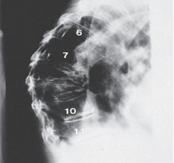
-Refer to the figure. What radiographic view is this?
A) AP
B) Lateral
C) Oblique
D) Coronal
سؤال
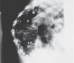
-Refer to the figure. The appearance of the osseous deformity infers what mechanism of injury?
A) Extension
B) Hyperextension
C) Flexion
D) Side-bending
سؤال
-The eighth and ninth vertebral deformity is associated with osteoporosis and the most likely diagnosis is:
A) Vertebral compression fractures
B) Tuberculosis of the spine
C) Herniated nucleus pulposus
D) Osteomyelitis of the spine
سؤال
-The radiologic evaluation of scoliosis uses the erect side-bending views to assess:
A) Bone age at the vertebral ring apophyses
B) Structural versus nonstructural curves
C) The Cobb value for the convexity
D) The Cobb value for the concavity
سؤال

-Refer to the figure. What imaging modality and view is this?
A) Lateral radiograph
B) Sagittal CT
C) Coronal MRI
D) Sagittal MRI
سؤال
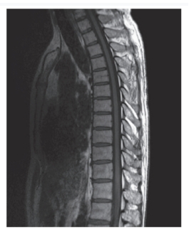
-Refer to the figure. Observe the spinal canal. The spinal canal would be described on this patient as:
A) Patent at all levels
B) Effaced at all levels
سؤال
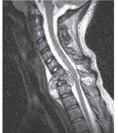
-Refer to the figure. What modality and sequence is this? Hint: Remember to look where you expect to see fluid.
A) T1-weighted CT
B) T2-weighted CT
C) T1-weighted MRI
D) T2-weighted MRI
سؤال

-Refer to the figure. What vertebrae has undergone complete destruction?
A) Seventh cervical vertebra
B) Eighth cervical vertebra
C) First thoracic vertebra
D) Second thoracic vertebra
سؤال
-In all age groups, __________________________of the vertebral bodies are the most common spinal injuries detected on radiographs.
A) herniated nucleus pulposus
B) anterior compression fracture
C) spondylolisthesis
D) Schmorl's nodes

فتح الحزمة
قم بالتسجيل لفتح البطاقات في هذه المجموعة!
Unlock Deck
Unlock Deck
1/10
العب
ملء الشاشة (f)
Deck 9: Radiologic Evaluation of the Thoracic Spine, Sternum, and Ribs
1
The most commonly fractured bones at the thoracic spine involve the:
A) Cervicothoracic region
B) Midthoracic region
C) Thoracolumbar region
D) Lumbosacral region
A) Cervicothoracic region
B) Midthoracic region
C) Thoracolumbar region
D) Lumbosacral region
Thoracolumbar region
2
-Refer to the figure. What radiographic view is this?
A) AP
B) Lateral
C) Oblique
D) Coronal
Lateral
3
-Refer to the figure. The appearance of the osseous deformity infers what mechanism of injury?
A) Extension
B) Hyperextension
C) Flexion
D) Side-bending
Flexion
4
-The eighth and ninth vertebral deformity is associated with osteoporosis and the most likely diagnosis is:
A) Vertebral compression fractures
B) Tuberculosis of the spine
C) Herniated nucleus pulposus
D) Osteomyelitis of the spine
فتح الحزمة
افتح القفل للوصول البطاقات البالغ عددها 10 في هذه المجموعة.
فتح الحزمة
k this deck
5
-The radiologic evaluation of scoliosis uses the erect side-bending views to assess:
A) Bone age at the vertebral ring apophyses
B) Structural versus nonstructural curves
C) The Cobb value for the convexity
D) The Cobb value for the concavity
فتح الحزمة
افتح القفل للوصول البطاقات البالغ عددها 10 في هذه المجموعة.
فتح الحزمة
k this deck
6
-Refer to the figure. What imaging modality and view is this?
A) Lateral radiograph
B) Sagittal CT
C) Coronal MRI
D) Sagittal MRI
فتح الحزمة
افتح القفل للوصول البطاقات البالغ عددها 10 في هذه المجموعة.
فتح الحزمة
k this deck
7
-Refer to the figure. Observe the spinal canal. The spinal canal would be described on this patient as:
A) Patent at all levels
B) Effaced at all levels
فتح الحزمة
افتح القفل للوصول البطاقات البالغ عددها 10 في هذه المجموعة.
فتح الحزمة
k this deck
8
-Refer to the figure. What modality and sequence is this? Hint: Remember to look where you expect to see fluid.
A) T1-weighted CT
B) T2-weighted CT
C) T1-weighted MRI
D) T2-weighted MRI
فتح الحزمة
افتح القفل للوصول البطاقات البالغ عددها 10 في هذه المجموعة.
فتح الحزمة
k this deck
9
-Refer to the figure. What vertebrae has undergone complete destruction?
A) Seventh cervical vertebra
B) Eighth cervical vertebra
C) First thoracic vertebra
D) Second thoracic vertebra
فتح الحزمة
افتح القفل للوصول البطاقات البالغ عددها 10 في هذه المجموعة.
فتح الحزمة
k this deck
10
-In all age groups, __________________________of the vertebral bodies are the most common spinal injuries detected on radiographs.
A) herniated nucleus pulposus
B) anterior compression fracture
C) spondylolisthesis
D) Schmorl's nodes
فتح الحزمة
افتح القفل للوصول البطاقات البالغ عددها 10 في هذه المجموعة.
فتح الحزمة
k this deck
فتح الحزمة
افتح القفل للوصول البطاقات البالغ عددها 10 في هذه المجموعة.


