Deck 7: Radiologic Evaluation of the Cervical Spine
ملء الشاشة (f)
سؤال
سؤال
سؤال
سؤال
سؤال

-Refer to the figure. Please identify the following view:
A) Anteroposterior
B) AP open mouth
C) AP lower cervical spine
D) Lateral
سؤال
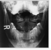
-Refer to the figure. What is most significant to identify on this view?
A) Position of the mandible
B) The C1-C2 disc space
C) Symmetry of the C1-C2 articulation
D) Cervical lordosis
سؤال
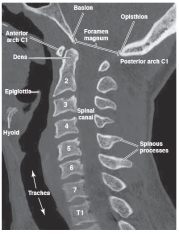
-Refer to the figure. What plane is this CT of the cervical spine?
A) Lateral
B) Sagittal
C) Coronal
D) Axial
سؤال

-Refer to the figure. The spinal canal is visible in its full anteroposterior diameter. Why is this possible?
A) This slice is reformatted at the midline
B) Bony pedicles and lamina are not well visualized on CT
C) The spinal cord has a greater density than the spinal canal
D) The spinal canal is only visible on an axial view.
سؤال

-Refer to the figure. This image is made in what plane?
A) Axial
B) Coronal
C) Sagittal
D) Oblique
سؤال
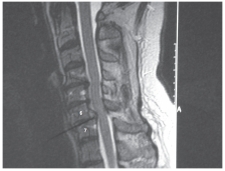
-Refer to the figure. The anterolisthesis of C6 on C7 has resulted in abnormal compression of the:
A) Spinal cord
B) Spinal nerve root
C) Intervertebral disk
D) Vertebral body

فتح الحزمة
قم بالتسجيل لفتح البطاقات في هذه المجموعة!
Unlock Deck
Unlock Deck
1/10
العب
ملء الشاشة (f)
Deck 7: Radiologic Evaluation of the Cervical Spine
1
Foraminal encroachment in the cervical spine would most likely result in which of the following symptoms?
A) Radiating arm pain
B) Cauda equina
C) Loss of proprioception
D) Neck stiffness
A) Radiating arm pain
B) Cauda equina
C) Loss of proprioception
D) Neck stiffness
Radiating arm pain
2
What radiographic view is used to screen for fracture or dislocation in the cervical spine?
A) Lateral flexion-extension view
B) Anteroposterior upper cervical spine view
C) Anteroposterior lower cervical spine view
D) Cross-table lateral view
A) Lateral flexion-extension view
B) Anteroposterior upper cervical spine view
C) Anteroposterior lower cervical spine view
D) Cross-table lateral view
Cross-table lateral view
3
Degenerative disk disease is seen on the lateral radiograph as a decrease in disk space height. On MRI it is characterized by:
A) Ossified discs
B) Dehydrated discs
C) High signal intensity
D) Inflammation of disks
A) Ossified discs
B) Dehydrated discs
C) High signal intensity
D) Inflammation of disks
Dehydrated discs
4
The hallmarks of degenerative joint disease in the cervical spine include decreased joint space, subchondral sclerosis, and osteophytosis of the:
A) Intervertebral disks
B) Vertebral bodies
C) Facet joints
D) Pedicles
A) Intervertebral disks
B) Vertebral bodies
C) Facet joints
D) Pedicles
فتح الحزمة
افتح القفل للوصول البطاقات البالغ عددها 10 في هذه المجموعة.
فتح الحزمة
k this deck
5
-Refer to the figure. Please identify the following view:
A) Anteroposterior
B) AP open mouth
C) AP lower cervical spine
D) Lateral
فتح الحزمة
افتح القفل للوصول البطاقات البالغ عددها 10 في هذه المجموعة.
فتح الحزمة
k this deck
6
-Refer to the figure. What is most significant to identify on this view?
A) Position of the mandible
B) The C1-C2 disc space
C) Symmetry of the C1-C2 articulation
D) Cervical lordosis
فتح الحزمة
افتح القفل للوصول البطاقات البالغ عددها 10 في هذه المجموعة.
فتح الحزمة
k this deck
7
-Refer to the figure. What plane is this CT of the cervical spine?
A) Lateral
B) Sagittal
C) Coronal
D) Axial
فتح الحزمة
افتح القفل للوصول البطاقات البالغ عددها 10 في هذه المجموعة.
فتح الحزمة
k this deck
8
-Refer to the figure. The spinal canal is visible in its full anteroposterior diameter. Why is this possible?
A) This slice is reformatted at the midline
B) Bony pedicles and lamina are not well visualized on CT
C) The spinal cord has a greater density than the spinal canal
D) The spinal canal is only visible on an axial view.
فتح الحزمة
افتح القفل للوصول البطاقات البالغ عددها 10 في هذه المجموعة.
فتح الحزمة
k this deck
9
-Refer to the figure. This image is made in what plane?
A) Axial
B) Coronal
C) Sagittal
D) Oblique
فتح الحزمة
افتح القفل للوصول البطاقات البالغ عددها 10 في هذه المجموعة.
فتح الحزمة
k this deck
10
-Refer to the figure. The anterolisthesis of C6 on C7 has resulted in abnormal compression of the:
A) Spinal cord
B) Spinal nerve root
C) Intervertebral disk
D) Vertebral body
فتح الحزمة
افتح القفل للوصول البطاقات البالغ عددها 10 في هذه المجموعة.
فتح الحزمة
k this deck
فتح الحزمة
افتح القفل للوصول البطاقات البالغ عددها 10 في هذه المجموعة.


