Deck 6: Diagnostic Ultrasound
ملء الشاشة (f)
سؤال
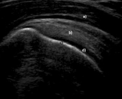
-Refer to the figure. What are the structures marked a-d in this normal longitudinal ultrasound of the shoulder?
A) (a) deltoid muscle, (b) subacromial bursa, (c) cancellous bone, (d) synovial fluid
B) (a) subacromial bursa, (b) supraspinatus muscle, (c) cortical bone, (d) articular cartilage
C) (a) subdeltoid bursa, (b) supraspinatus tendon, (c) cancellous, (d) articular capsule
D) (a) deltoid muscle, (b) supraspinatus tendon, (c) cortical bone, (d) articular cartilage
سؤال
سؤال
سؤال
سؤال
سؤال
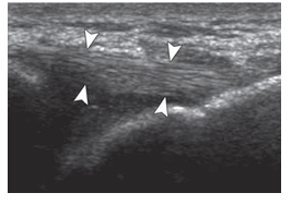
-Refer to the figure. What is the structure between the arrowheads?
A) Muscle
B) Ligament
C) Bursa
D) Subcutaneous fat
سؤال
سؤال
سؤال
سؤال

-Refer to the figure. If you compare the longitudinal ultrasound scan of the supraspinatus muscle and tendon (arrowheads) on the right with the normal supraspinatus on the left, what are your findings?
A) Thickened supraspinatus tendon
B) Swollen subacromial bursa
C) Cortical irregularities and a partial tear
D) Hyperechoic focus in the distal tendon

فتح الحزمة
قم بالتسجيل لفتح البطاقات في هذه المجموعة!
Unlock Deck
Unlock Deck
1/10
العب
ملء الشاشة (f)
Deck 6: Diagnostic Ultrasound
1
-Refer to the figure. What are the structures marked a-d in this normal longitudinal ultrasound of the shoulder?
A) (a) deltoid muscle, (b) subacromial bursa, (c) cancellous bone, (d) synovial fluid
B) (a) subacromial bursa, (b) supraspinatus muscle, (c) cortical bone, (d) articular cartilage
C) (a) subdeltoid bursa, (b) supraspinatus tendon, (c) cancellous, (d) articular capsule
D) (a) deltoid muscle, (b) supraspinatus tendon, (c) cortical bone, (d) articular cartilage
(a) deltoid muscle, (b) supraspinatus tendon, (c) cortical bone, (d) articular cartilage
2
What are the differences between linear and curvilinear transducers?
A) The linear transducer is often used for abdominopelvic ultrasound
B) The linear transducer displays a field of view wider than the transducer itself
C) The linear transducer is predominantly used for musculoskeletal imaging
D) A curved transducer displays a wider field of view for structures that are close to the transducer
The linear transducer is predominantly used for musculoskeletal imaging
3
High signal intensity in ultrasound images is seen when there is:
A) High absorption of ultrasound
B) Reflection of ultrasound waves tissue off interfaces with similar impedance
AU: Is a word missing here between waves and tissue?
It should read "…ultrasound waves off tissue".
C) Scattering of the ultrasound energy
D) Ultrasound is reflected off smooth surfaces
A) High absorption of ultrasound
B) Reflection of ultrasound waves tissue off interfaces with similar impedance
AU: Is a word missing here between waves and tissue?
It should read "…ultrasound waves off tissue".
C) Scattering of the ultrasound energy
D) Ultrasound is reflected off smooth surfaces
Ultrasound is reflected off smooth surfaces
4
In Doppler ultrasound, a red color indicates:
A) A high velocity of blood flow toward the transducer
B) Irregular blood flow in a tortuous vessel
C) Slow blood flow
D) A small blood vessel
A) A high velocity of blood flow toward the transducer
B) Irregular blood flow in a tortuous vessel
C) Slow blood flow
D) A small blood vessel
فتح الحزمة
افتح القفل للوصول البطاقات البالغ عددها 10 في هذه المجموعة.
فتح الحزمة
k this deck
5
Order these tissues from lowest to highest ultrasound signal intensity:
A) Bone, tendon, muscle, hyaline cartilage
B) Hyaline cartilage, bone, tendon, muscle
C) Hyaline cartilage, muscle, tendon, bone
D) Muscle, hyaline cartilage, tendon, bone
A) Bone, tendon, muscle, hyaline cartilage
B) Hyaline cartilage, bone, tendon, muscle
C) Hyaline cartilage, muscle, tendon, bone
D) Muscle, hyaline cartilage, tendon, bone
فتح الحزمة
افتح القفل للوصول البطاقات البالغ عددها 10 في هذه المجموعة.
فتح الحزمة
k this deck
6
-Refer to the figure. What is the structure between the arrowheads?
A) Muscle
B) Ligament
C) Bursa
D) Subcutaneous fat
فتح الحزمة
افتح القفل للوصول البطاقات البالغ عددها 10 في هذه المجموعة.
فتح الحزمة
k this deck
7
The scanning planes in musculoskeletal ultrasound can best be described as:
A) Comparable to CT and MRI
B) Transverse, when viewing muscles
C) A direct continuation of the transducer orientation
D) An extended field of view, when looking at a muscle longitudinally
A) Comparable to CT and MRI
B) Transverse, when viewing muscles
C) A direct continuation of the transducer orientation
D) An extended field of view, when looking at a muscle longitudinally
فتح الحزمة
افتح القفل للوصول البطاقات البالغ عددها 10 في هذه المجموعة.
فتح الحزمة
k this deck
8
Which is true of the imaging characteristics of an injured tendon?
A) Strains are displayed as increased signal intensity
B) It shows a hyperechoic, parallel fiber pattern
C) Strains show thinning of the tendon
D) The site of a tendon rupture may initially be hypoechoic
A) Strains are displayed as increased signal intensity
B) It shows a hyperechoic, parallel fiber pattern
C) Strains show thinning of the tendon
D) The site of a tendon rupture may initially be hypoechoic
فتح الحزمة
افتح القفل للوصول البطاقات البالغ عددها 10 في هذه المجموعة.
فتح الحزمة
k this deck
9
The advantages of ultrasound imaging include all except:
A) Wide field of view
B) Higher resolution
C) Ability to modify the imaging while it is being done
D) Lower cost
A) Wide field of view
B) Higher resolution
C) Ability to modify the imaging while it is being done
D) Lower cost
فتح الحزمة
افتح القفل للوصول البطاقات البالغ عددها 10 في هذه المجموعة.
فتح الحزمة
k this deck
10
-Refer to the figure. If you compare the longitudinal ultrasound scan of the supraspinatus muscle and tendon (arrowheads) on the right with the normal supraspinatus on the left, what are your findings?
A) Thickened supraspinatus tendon
B) Swollen subacromial bursa
C) Cortical irregularities and a partial tear
D) Hyperechoic focus in the distal tendon
فتح الحزمة
افتح القفل للوصول البطاقات البالغ عددها 10 في هذه المجموعة.
فتح الحزمة
k this deck
فتح الحزمة
افتح القفل للوصول البطاقات البالغ عددها 10 في هذه المجموعة.


