Deck 8: Presumptive Identification
ملء الشاشة (f)
سؤال
A microbiologist performs a Gram stain on a positive blood culture bottle. She does not observe any organisms. She then uses an acridine orange stain and observes the organisms pictured in the image. What type of microscope did she use to observe the rod-like organisms?
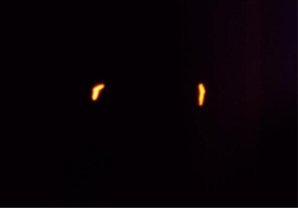
A) Brightfield
B) Darkfield
C) Phase contrast
D) Fluorescent
A) Brightfield
B) Darkfield
C) Phase contrast
D) Fluorescent
سؤال
This image of the spirochete Treponema pallidum was photographed using a _____________ microscope.
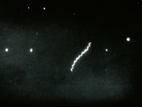
A) brightfield
B) darkfield
C) phase contrast
D) fluorescent
A) brightfield
B) darkfield
C) phase contrast
D) fluorescent
سؤال
سؤال
سؤال
سؤال
سؤال
سؤال
سؤال
سؤال
سؤال
سؤال
سؤال
سؤال
Review the Gram stain image of a cerebrospinal fluid. Based on the results, you should
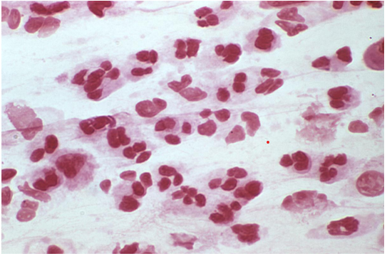
A) look at a drop of the specimen under a darkfield microscope.
B) prepare a new slide and perform a Kinyoun stain.
C) prepare a new slide and perform an acridine orange stain.
D) report the slide as "no organisms seen."
A) look at a drop of the specimen under a darkfield microscope.
B) prepare a new slide and perform a Kinyoun stain.
C) prepare a new slide and perform an acridine orange stain.
D) report the slide as "no organisms seen."
سؤال
Review the Gram stain image provided of a positive blood culture bottle. It should be
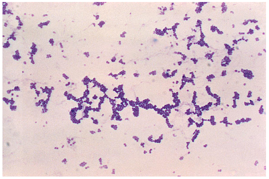
A) pleomorphic gram-positive coccobacilli.
B) gram-positive cocci in pairs and chains.
C) gram-positive cocci in clusters.
D) palisading gram-negative cocci.
A) pleomorphic gram-positive coccobacilli.
B) gram-positive cocci in pairs and chains.
C) gram-positive cocci in clusters.
D) palisading gram-negative cocci.
سؤال
Review the image of a leg wound culture provided. The organisms appear as gram-variable rods. What Gram stain reaction should be reported for this organism?
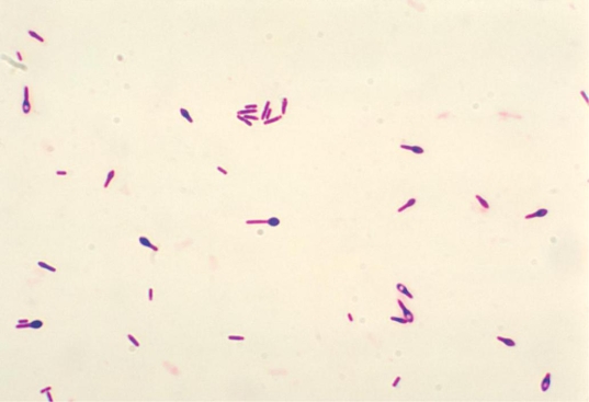 Source: CDC and Dr. George Lombard
A) Gram negative
B) Gram positive
C) Gram variable
D) Gram positive and gram negative
Source: CDC and Dr. George LombardA) Gram negative
B) Gram positive
C) Gram variable
D) Gram positive and gram negative
سؤال
Based on the colonies observed in the image provided, you can deduce the organism
1) is a gram-negative rod.
2) is a gram-positive coccus.
3) produces a capsule.
4) produces a pigment.
A) 1, 3, and 4
B) 2, 3, and 4
C) 2 and 4
D) 1 and 3
1) is a gram-negative rod.
2) is a gram-positive coccus.
3) produces a capsule.
4) produces a pigment.
A) 1, 3, and 4
B) 2, 3, and 4
C) 2 and 4
D) 1 and 3
سؤال
سؤال
Review the Gram stained urine image provided. Which organism listed best correlates with this Gram stain reaction?
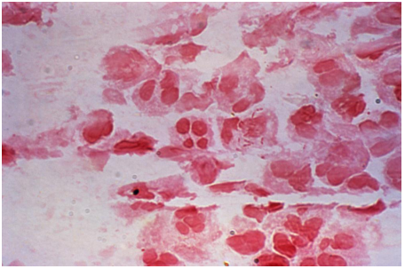
A) Beta-hemolytic, catalase negative, and PYR positive
B) Beta-hemolytic, catalase and coagulase positive
C) Gamma-hemolytic, swarmer, oxidase, and spot indole negative
D) Alpha-hemolytic, catalase negative, and bile soluble
A) Beta-hemolytic, catalase negative, and PYR positive
B) Beta-hemolytic, catalase and coagulase positive
C) Gamma-hemolytic, swarmer, oxidase, and spot indole negative
D) Alpha-hemolytic, catalase negative, and bile soluble
سؤال

فتح الحزمة
قم بالتسجيل لفتح البطاقات في هذه المجموعة!
Unlock Deck
Unlock Deck
1/20
العب
ملء الشاشة (f)
Deck 8: Presumptive Identification
1
A microbiologist performs a Gram stain on a positive blood culture bottle. She does not observe any organisms. She then uses an acridine orange stain and observes the organisms pictured in the image. What type of microscope did she use to observe the rod-like organisms?
A) Brightfield
B) Darkfield
C) Phase contrast
D) Fluorescent
A) Brightfield
B) Darkfield
C) Phase contrast
D) Fluorescent
Fluorescent
2
This image of the spirochete Treponema pallidum was photographed using a _____________ microscope.
A) brightfield
B) darkfield
C) phase contrast
D) fluorescent
A) brightfield
B) darkfield
C) phase contrast
D) fluorescent
darkfield
3
Which of the following microscopes requires that the specimen be stained to observe microorganisms and cells?
A) Fluorescent
B) Phase contrast
C) Darkfield
D) Electron
A) Fluorescent
B) Phase contrast
C) Darkfield
D) Electron
Fluorescent
4
The eyepiece of a brightfield microscope contains the ___________ lens.
A) objective
B) ocular
C) condenser
D) diaphragm
A) objective
B) ocular
C) condenser
D) diaphragm
فتح الحزمة
افتح القفل للوصول البطاقات البالغ عددها 20 في هذه المجموعة.
فتح الحزمة
k this deck
5
All of the following play a role in setting Köhler illumination of a brightfield microscope except
A) stage.
B) substage condenser.
C) field diaphragm.
D) iris diaphragm.
A) stage.
B) substage condenser.
C) field diaphragm.
D) iris diaphragm.
فتح الحزمة
افتح القفل للوصول البطاقات البالغ عددها 20 في هذه المجموعة.
فتح الحزمة
k this deck
6
The purpose of Köhler illumination is to
A) ensure adequate resolution.
B) ensure uniform illumination.
C) neither a nor b.
D) both a and b.
A) ensure adequate resolution.
B) ensure uniform illumination.
C) neither a nor b.
D) both a and b.
فتح الحزمة
افتح القفل للوصول البطاقات البالغ عددها 20 في هذه المجموعة.
فتح الحزمة
k this deck
7
Setting a brightfield microscope up for Köhler illumination must be repeated for each
A) slide read.
B) day of use.
C) objective.
D) co-worker.
A) slide read.
B) day of use.
C) objective.
D) co-worker.
فتح الحزمة
افتح القفل للوصول البطاقات البالغ عددها 20 في هذه المجموعة.
فتح الحزمة
k this deck
8
At the end of the day, you are the last to use the brightfield microscope. Before leaving you will
A) wipe the microscope down.
B) remove oil from the 100X objective.
C) reset it for Köhler illumination.
D) leave it as is for the next shift.
A) wipe the microscope down.
B) remove oil from the 100X objective.
C) reset it for Köhler illumination.
D) leave it as is for the next shift.
فتح الحزمة
افتح القفل للوصول البطاقات البالغ عددها 20 في هذه المجموعة.
فتح الحزمة
k this deck
9
A brightfield microscope should be thoroughly maintained at least
A) weekly.
B) monthly.
C) biannually.
D) annually.
A) weekly.
B) monthly.
C) biannually.
D) annually.
فتح الحزمة
افتح القفل للوصول البطاقات البالغ عددها 20 في هذه المجموعة.
فتح الحزمة
k this deck
10
You cannot find material on a Gram stained cerebrospinal fluid slide to focus on using the 10X objective. You should
A) turn the slide over. It is upside down.
B) reset the microscope for Köhler illumination and refocus.
C) raise the condenser to allow more light on the slide.
D) focus on the frosted end of the slide and then move to the specimen area.
A) turn the slide over. It is upside down.
B) reset the microscope for Köhler illumination and refocus.
C) raise the condenser to allow more light on the slide.
D) focus on the frosted end of the slide and then move to the specimen area.
فتح الحزمة
افتح القفل للوصول البطاقات البالغ عددها 20 في هذه المجموعة.
فتح الحزمة
k this deck
11
The light bulb on the brightfield microscope burns out. You replace it and it burns out again when you turn on the light. This can be due to
A) electrical surge from the outlet.
B) incorrect bulb wattage.
C) rheostat that is set too high.
D) broken electrical cord.
A) electrical surge from the outlet.
B) incorrect bulb wattage.
C) rheostat that is set too high.
D) broken electrical cord.
فتح الحزمة
افتح القفل للوصول البطاقات البالغ عددها 20 في هذه المجموعة.
فتح الحزمة
k this deck
12
Select the correct combination of stain and microscope to detect the capsule of the yeast Cryptococcus neoformans.
A)
B)
C)
D)
A)
B)
C)
D)
فتح الحزمة
افتح القفل للوصول البطاقات البالغ عددها 20 في هذه المجموعة.
فتح الحزمة
k this deck
13
The acid fast stains Ziehl-Neelsen and Kinyoun can be differentiated by the
A) use of heat to facilitate stain penetration in the Ziehl-Neelsen method.
B) use of heat to facilitate stain penetration in the Kinyoun method.
C) the use of carbolfuchsin for the Ziehl-Neelsen method.
D) the use of carbolfuchsin for the Kinyoun method.
A) use of heat to facilitate stain penetration in the Ziehl-Neelsen method.
B) use of heat to facilitate stain penetration in the Kinyoun method.
C) the use of carbolfuchsin for the Ziehl-Neelsen method.
D) the use of carbolfuchsin for the Kinyoun method.
فتح الحزمة
افتح القفل للوصول البطاقات البالغ عددها 20 في هذه المجموعة.
فتح الحزمة
k this deck
14
Review the Gram stain image of a cerebrospinal fluid. Based on the results, you should
A) look at a drop of the specimen under a darkfield microscope.
B) prepare a new slide and perform a Kinyoun stain.
C) prepare a new slide and perform an acridine orange stain.
D) report the slide as "no organisms seen."
A) look at a drop of the specimen under a darkfield microscope.
B) prepare a new slide and perform a Kinyoun stain.
C) prepare a new slide and perform an acridine orange stain.
D) report the slide as "no organisms seen."
فتح الحزمة
افتح القفل للوصول البطاقات البالغ عددها 20 في هذه المجموعة.
فتح الحزمة
k this deck
15
Review the Gram stain image provided of a positive blood culture bottle. It should be
A) pleomorphic gram-positive coccobacilli.
B) gram-positive cocci in pairs and chains.
C) gram-positive cocci in clusters.
D) palisading gram-negative cocci.
A) pleomorphic gram-positive coccobacilli.
B) gram-positive cocci in pairs and chains.
C) gram-positive cocci in clusters.
D) palisading gram-negative cocci.
فتح الحزمة
افتح القفل للوصول البطاقات البالغ عددها 20 في هذه المجموعة.
فتح الحزمة
k this deck
16
Review the image of a leg wound culture provided. The organisms appear as gram-variable rods. What Gram stain reaction should be reported for this organism?
Source: CDC and Dr. George Lombard
A) Gram negative
B) Gram positive
C) Gram variable
D) Gram positive and gram negative
Source: CDC and Dr. George LombardA) Gram negative
B) Gram positive
C) Gram variable
D) Gram positive and gram negative
فتح الحزمة
افتح القفل للوصول البطاقات البالغ عددها 20 في هذه المجموعة.
فتح الحزمة
k this deck
17
Based on the colonies observed in the image provided, you can deduce the organism
1) is a gram-negative rod.
2) is a gram-positive coccus.
3) produces a capsule.
4) produces a pigment.
A) 1, 3, and 4
B) 2, 3, and 4
C) 2 and 4
D) 1 and 3
1) is a gram-negative rod.
2) is a gram-positive coccus.
3) produces a capsule.
4) produces a pigment.
A) 1, 3, and 4
B) 2, 3, and 4
C) 2 and 4
D) 1 and 3
فتح الحزمة
افتح القفل للوصول البطاقات البالغ عددها 20 في هذه المجموعة.
فتح الحزمة
k this deck
18
Partially lysed red blood cells under and around a colony on sheep blood agar appear as
A) no change in the agar.
B) a clear zone.
C) a green discoloration.
D) a blue discoloration.
A) no change in the agar.
B) a clear zone.
C) a green discoloration.
D) a blue discoloration.
فتح الحزمة
افتح القفل للوصول البطاقات البالغ عددها 20 في هذه المجموعة.
فتح الحزمة
k this deck
19
Review the Gram stained urine image provided. Which organism listed best correlates with this Gram stain reaction?
A) Beta-hemolytic, catalase negative, and PYR positive
B) Beta-hemolytic, catalase and coagulase positive
C) Gamma-hemolytic, swarmer, oxidase, and spot indole negative
D) Alpha-hemolytic, catalase negative, and bile soluble
A) Beta-hemolytic, catalase negative, and PYR positive
B) Beta-hemolytic, catalase and coagulase positive
C) Gamma-hemolytic, swarmer, oxidase, and spot indole negative
D) Alpha-hemolytic, catalase negative, and bile soluble
فتح الحزمة
افتح القفل للوصول البطاقات البالغ عددها 20 في هذه المجموعة.
فتح الحزمة
k this deck
20
A positive blood culture vial reveals gram-negative rods. After subculture, the organism grew as
Sheep blood agar: large, gray colonies with beta-hemolysis
MacConkey agar: round, smooth, pink colonies
Phenylethyl alcohol agar: no growth
Rapid testing revealed
Oxidase: no color change
Pigment: none observed
Spot indole: red-pink
The organism is
A) Escherichia coli.
B) Pseudomonas aeruginosa.
C) Proteus mirabilis.
D) Proteus vulgaris.
فتح الحزمة
افتح القفل للوصول البطاقات البالغ عددها 20 في هذه المجموعة.
فتح الحزمة
k this deck
فتح الحزمة
افتح القفل للوصول البطاقات البالغ عددها 20 في هذه المجموعة.


