Deck 20: Skull, Facial Bones, and Paranasal Sinuses
ملء الشاشة (f)
سؤال
سؤال
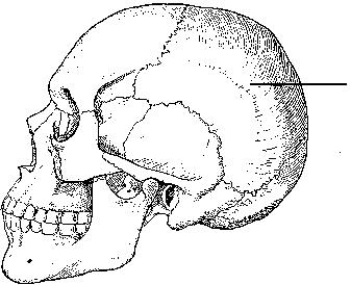
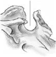
The part of the frontal bone indicated in the figure below is the:
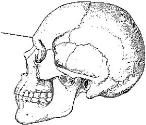
A) bregma.
B) lambda.
C) glabella.
D) acanthion.
A) bregma.
B) lambda.
C) glabella.
D) acanthion.
سؤال
سؤال
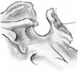
The bone indicated in the figure below is the:
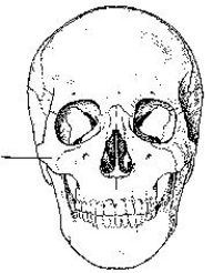
A) parietal.
B) orbital.
C) temporal.
D) zygoma.
A) parietal.
B) orbital.
C) temporal.
D) zygoma.
سؤال
The bone identified in the figure below is the:
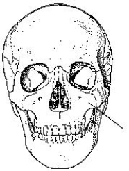
A) maxilla.
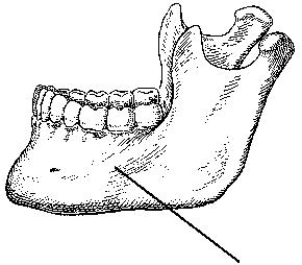
B) frontal.
C) mandible.
D) ethmoid.
A) maxilla.
B) frontal.
C) mandible.
D) ethmoid.
سؤال
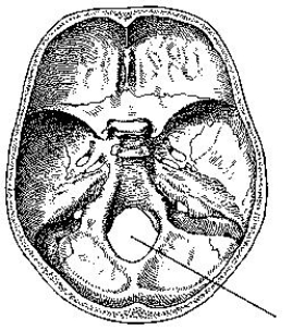
The part of the cranial base identified in the figure below is the:
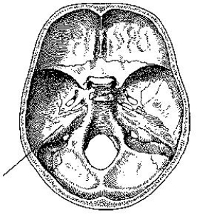
A) auditory canal.
B) dorsum sellae.
C) greater wing.
D) petrous portion.
A) auditory canal.
B) dorsum sellae.
C) greater wing.
D) petrous portion.
سؤال
The bone indicated in the figure below is the:
A) temporal.
B) parietal.
C) occipital.
D) sphenoid.
A) temporal.
B) parietal.
C) occipital.
D) sphenoid.
سؤال
سؤال
سؤال
The part of the cranial base identified in the figure below is the:
A) sella turcica.
B) foramen ovale.
C) hypoglossal canal.
D) foramen magnum.
A) sella turcica.
B) foramen ovale.
C) hypoglossal canal.
D) foramen magnum.
سؤال
سؤال
سؤال
سؤال
سؤال
سؤال
سؤال
سؤال
سؤال
سؤال
سؤال
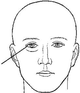
The landmark identified in the figure below is termed the:
A) nasion.
B) glabella.
C) acanthion.
D) inner canthus.
A) nasion.
B) glabella.
C) acanthion.
D) inner canthus.
سؤال
سؤال
The external landmark identified in the figure below is the:
A) glabella.
B) acanthion.
C) outer canthus.
D) infraorbital margin.
A) glabella.
B) acanthion.
C) outer canthus.
D) infraorbital margin.
سؤال
سؤال
سؤال
سؤال
The part of the mandible identified in the figure below is the:
A) body.
B) ramus.
C) symphysis.
D) alveolar portion.
A) body.
B) ramus.
C) symphysis.
D) alveolar portion.
سؤال
The portion of the mandible identified in the figure below is the:
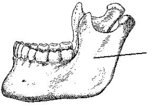
A) body.
B) ramus.
C) symphysis.
D) alveolar portion.
A) body.
B) ramus.
C) symphysis.
D) alveolar portion.
سؤال
سؤال
سؤال
The landmark identified in the figure below is the:
A) nasion.
B) glabella.
C) acanthion.
D) auricular point.
A) nasion.
B) glabella.
C) acanthion.
D) auricular point.
سؤال
سؤال
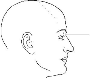
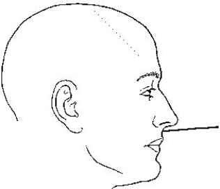
The topographic line shown in the figure below is the _____ line.
A) glabellomeatal
B) orbitomeatal
C) acanthiomeatal
D) infraorbitomeatal
A) glabellomeatal
B) orbitomeatal
C) acanthiomeatal
D) infraorbitomeatal
سؤال
سؤال
سؤال
سؤال
سؤال
سؤال
سؤال
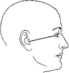
The topographic line identified in the figure below is the _____ line.
A) orbitomeatal
B) mentomeatal
C) acanthiomeatal
D) infraorbitomeatal
A) orbitomeatal
B) mentomeatal
C) acanthiomeatal
D) infraorbitomeatal
سؤال
سؤال
سؤال
سؤال
Which method of examining the skull is identified in the figure below?
A) Haas
B) Towne
C) Shüller
D) Caldwell
A) Haas
B) Towne
C) Shüller
D) Caldwell
سؤال
سؤال
سؤال
سؤال
سؤال
سؤال
سؤال
سؤال
سؤال
سؤال
سؤال
سؤال
سؤال
سؤال
سؤال
سؤال
The x-ray projection demonstrated in the figure below is the:
A) SMV.
B) VSM.
C) AP axial.
D) PA axial.
A) SMV.
B) VSM.
C) AP axial.
D) PA axial.
سؤال
سؤال
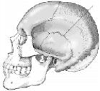
The bone identified on the skull below is the:
A) temporal.
B) parietal.
C) zygoma.
D) sphenoid.
A) temporal.
B) parietal.
C) zygoma.
D) sphenoid.
سؤال
سؤال
The part of the sphenoid bone identified in the figure below is the:
A) clivus.
B) foramen magnum.
C) sella turcica.
D) dorsum sellae.
A) clivus.
B) foramen magnum.
C) sella turcica.
D) dorsum sellae.
سؤال
سؤال
سؤال
سؤال
The part of the sphenoid bone identified in the figure below is the:
A) clivus.
B) clinoid processes.
C) sella turcica.
D) dorsum sellae.
A) clivus.
B) clinoid processes.
C) sella turcica.
D) dorsum sellae.
سؤال
سؤال
سؤال
سؤال
سؤال
سؤال
سؤال
سؤال
The suture identified on the figure below is the:
A) coronal.
B) squamosal.
C) sagittal.
D) lambdoidal.
A) coronal.
B) squamosal.
C) sagittal.
D) lambdoidal.
سؤال
سؤال
سؤال
The bone identified on the skull below is the:
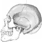
A) temporal.
B) parietal.
C) occipital.
D) sphenoid.
A) temporal.
B) parietal.
C) occipital.
D) sphenoid.
سؤال

فتح الحزمة
قم بالتسجيل لفتح البطاقات في هذه المجموعة!
Unlock Deck
Unlock Deck
1/196
العب
ملء الشاشة (f)
Deck 20: Skull, Facial Bones, and Paranasal Sinuses
1
All of the following are cranial bones except the:
A) maxillae.
B) frontal.
C) sphenoid.
D) occipital.
A) maxillae.
B) frontal.
C) sphenoid.
D) occipital.
maxillae.
2
The part of the frontal bone indicated in the figure below is the:
A) bregma.
B) lambda.
C) glabella.
D) acanthion.
A) bregma.
B) lambda.
C) glabella.
D) acanthion.
glabella.
3
All of the following are facial bones except the:
A) ethmoid.
B) maxillae.
C) mandible.
D) zygomatic bones.
A) ethmoid.
B) maxillae.
C) mandible.
D) zygomatic bones.
ethmoid.
4
The bone indicated in the figure below is the:
A) parietal.
B) orbital.
C) temporal.
D) zygoma.
A) parietal.
B) orbital.
C) temporal.
D) zygoma.
فتح الحزمة
افتح القفل للوصول البطاقات البالغ عددها 196 في هذه المجموعة.
فتح الحزمة
k this deck
5
The bone identified in the figure below is the:
A) maxilla.
B) frontal.
C) mandible.
D) ethmoid.
A) maxilla.
B) frontal.
C) mandible.
D) ethmoid.
فتح الحزمة
افتح القفل للوصول البطاقات البالغ عددها 196 في هذه المجموعة.
فتح الحزمة
k this deck
6
The part of the cranial base identified in the figure below is the:
A) auditory canal.
B) dorsum sellae.
C) greater wing.
D) petrous portion.
A) auditory canal.
B) dorsum sellae.
C) greater wing.
D) petrous portion.
فتح الحزمة
افتح القفل للوصول البطاقات البالغ عددها 196 في هذه المجموعة.
فتح الحزمة
k this deck
7
The bone indicated in the figure below is the:
A) temporal.
B) parietal.
C) occipital.
D) sphenoid.
A) temporal.
B) parietal.
C) occipital.
D) sphenoid.
فتح الحزمة
افتح القفل للوصول البطاقات البالغ عددها 196 في هذه المجموعة.
فتح الحزمة
k this deck
8
Which bone in the skull contains the auditory organs and the organs of hearing?
A) Temporal
B) Sphenoid
C) Occipital
D) Ethmoid
A) Temporal
B) Sphenoid
C) Occipital
D) Ethmoid
فتح الحزمة
افتح القفل للوصول البطاقات البالغ عددها 196 في هذه المجموعة.
فتح الحزمة
k this deck
9
Which of the following is located in the middle ear?
A) Cochlea
B) Bony labyrinth
C) Tympanic membrane
D) External acoustic meatus
A) Cochlea
B) Bony labyrinth
C) Tympanic membrane
D) External acoustic meatus
فتح الحزمة
افتح القفل للوصول البطاقات البالغ عددها 196 في هذه المجموعة.
فتح الحزمة
k this deck
10
The part of the cranial base identified in the figure below is the:
A) sella turcica.
B) foramen ovale.
C) hypoglossal canal.
D) foramen magnum.
A) sella turcica.
B) foramen ovale.
C) hypoglossal canal.
D) foramen magnum.
فتح الحزمة
افتح القفل للوصول البطاقات البالغ عددها 196 في هذه المجموعة.
فتح الحزمة
k this deck
11
How many bones make up the cranium?
A) 4
B) 6
C) 8
D) 10
A) 4
B) 6
C) 8
D) 10
فتح الحزمة
افتح القفل للوصول البطاقات البالغ عددها 196 في هذه المجموعة.
فتح الحزمة
k this deck
12
The petromastoid portion is a part of which bone?
A) Temporal
B) Sphenoid
C) Occipital
D) Ethmoid
A) Temporal
B) Sphenoid
C) Occipital
D) Ethmoid
فتح الحزمة
افتح القفل للوصول البطاقات البالغ عددها 196 في هذه المجموعة.
فتح الحزمة
k this deck
13
The zygomatic arches are a part of which bone?
A) Frontal
B) Parietal
C) Temporal
D) Sphenoid
A) Frontal
B) Parietal
C) Temporal
D) Sphenoid
فتح الحزمة
افتح القفل للوصول البطاقات البالغ عددها 196 في هذه المجموعة.
فتح الحزمة
k this deck
14
Which skull suture is found between the frontal and parietal bones?
A) Sagittal
B) Coronal
C) Squamosal
D) Lambdoidal
A) Sagittal
B) Coronal
C) Squamosal
D) Lambdoidal
فتح الحزمة
افتح القفل للوصول البطاقات البالغ عددها 196 في هذه المجموعة.
فتح الحزمة
k this deck
15
The vestibulocochlear organ is the organ of:
1)hearing.
2)sensation.
3)balance.
A) 1 and 2
B) 1 and 3
C) 2 and 3
D) 1, 2, and 3
1)hearing.
2)sensation.
3)balance.
A) 1 and 2
B) 1 and 3
C) 2 and 3
D) 1, 2, and 3
فتح الحزمة
افتح القفل للوصول البطاقات البالغ عددها 196 في هذه المجموعة.
فتح الحزمة
k this deck
16
Which skull suture is located between the parietal bones?
A) Hyoid
B) Coronal
C) Sagittal
D) Squamosal
A) Hyoid
B) Coronal
C) Sagittal
D) Squamosal
فتح الحزمة
افتح القفل للوصول البطاقات البالغ عددها 196 في هذه المجموعة.
فتح الحزمة
k this deck
17
How many bones make up the face?
A) 6
B) 10
C) 12
D) 14
A) 6
B) 10
C) 12
D) 14
فتح الحزمة
افتح القفل للوصول البطاقات البالغ عددها 196 في هذه المجموعة.
فتح الحزمة
k this deck
18
Which of the following bones contain air sinuses?
1)Ethmoid
2)Frontal
3)Sphenoid
A) 1 and 2
B) 1 and 3
C) 2 and 3
D) 1, 2, and 3
1)Ethmoid
2)Frontal
3)Sphenoid
A) 1 and 2
B) 1 and 3
C) 2 and 3
D) 1, 2, and 3
فتح الحزمة
افتح القفل للوصول البطاقات البالغ عددها 196 في هذه المجموعة.
فتح الحزمة
k this deck
19
The cranial bones are rigidly jointed together by articulations called:
A) joints.
B) bursae.
C) sutures.
D) cartilage.
A) joints.
B) bursae.
C) sutures.
D) cartilage.
فتح الحزمة
افتح القفل للوصول البطاقات البالغ عددها 196 في هذه المجموعة.
فتح الحزمة
k this deck
20
Which bone has condyles that articulate with the atlas of the cervical spine?
A) Temporal
B) Occipital
C) Parietal
D) Foramen magnum
A) Temporal
B) Occipital
C) Parietal
D) Foramen magnum
فتح الحزمة
افتح القفل للوصول البطاقات البالغ عددها 196 في هذه المجموعة.
فتح الحزمة
k this deck
21
The landmark identified in the figure below is termed the:
A) nasion.
B) glabella.
C) acanthion.
D) inner canthus.
A) nasion.
B) glabella.
C) acanthion.
D) inner canthus.
فتح الحزمة
افتح القفل للوصول البطاقات البالغ عددها 196 في هذه المجموعة.
فتح الحزمة
k this deck
22
The central-ray angle for the PA axial (Caldwell) projection of the skull is:
A) 5 degrees cephalad.
B) 10 degrees cephalad.
C) 12 degrees caudad.
D) 15 degrees caudad.
A) 5 degrees cephalad.
B) 10 degrees cephalad.
C) 12 degrees caudad.
D) 15 degrees caudad.
فتح الحزمة
افتح القفل للوصول البطاقات البالغ عددها 196 في هذه المجموعة.
فتح الحزمة
k this deck
23
The external landmark identified in the figure below is the:
A) glabella.
B) acanthion.
C) outer canthus.
D) infraorbital margin.
A) glabella.
B) acanthion.
C) outer canthus.
D) infraorbital margin.
فتح الحزمة
افتح القفل للوصول البطاقات البالغ عددها 196 في هذه المجموعة.
فتح الحزمة
k this deck
24
Which of the following should be seen superimposed on a lateral image of the skull?
1)Orbital roofs
2)External acoustic meati
3)Temporomandibular joints
A) 1 and 2
B) 1 and 3
C) 2 and 3
D) 1, 2, and 3
1)Orbital roofs
2)External acoustic meati
3)Temporomandibular joints
A) 1 and 2
B) 1 and 3
C) 2 and 3
D) 1, 2, and 3
فتح الحزمة
افتح القفل للوصول البطاقات البالغ عددها 196 في هذه المجموعة.
فتح الحزمة
k this deck
25
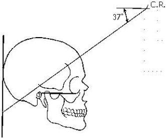
In a typically shaped head, the petrous pyramids project anteriorly and medially at what angle?
A) 37 degrees
B) 40 degrees
C) 47 degrees
D) 54 degrees
A) 37 degrees
B) 40 degrees
C) 47 degrees
D) 54 degrees
فتح الحزمة
افتح القفل للوصول البطاقات البالغ عددها 196 في هذه المجموعة.
فتح الحزمة
k this deck
26
The small bone situated at the base of the tongue is the:
A) hyoid.
B) alveolar.
C) cornu.
D) styloid.
A) hyoid.
B) alveolar.
C) cornu.
D) styloid.
فتح الحزمة
افتح القفل للوصول البطاقات البالغ عددها 196 في هذه المجموعة.
فتح الحزمة
k this deck
27
The part of the mandible identified in the figure below is the:
A) body.
B) ramus.
C) symphysis.
D) alveolar portion.
A) body.
B) ramus.
C) symphysis.
D) alveolar portion.
فتح الحزمة
افتح القفل للوصول البطاقات البالغ عددها 196 في هذه المجموعة.
فتح الحزمة
k this deck
28
The portion of the mandible identified in the figure below is the:
A) body.
B) ramus.
C) symphysis.
D) alveolar portion.
A) body.
B) ramus.
C) symphysis.
D) alveolar portion.
فتح الحزمة
افتح القفل للوصول البطاقات البالغ عددها 196 في هذه المجموعة.
فتح الحزمة
k this deck
29
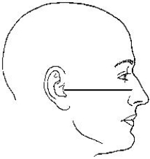
The central ray and center of the image receptor position for a lateral projection of the skull is _____ inch(es) _____ the EAM.
A) 1; below
B) 2; below
C) 1; above
D) 2; above
A) 1; below
B) 2; below
C) 1; above
D) 2; above
فتح الحزمة
افتح القفل للوصول البطاقات البالغ عددها 196 في هذه المجموعة.
فتح الحزمة
k this deck
30
Which of the following is located in the internal ear?
A) Concha
B) Auditory tube
C) Tympanic membrane
D) Semicircular canals
A) Concha
B) Auditory tube
C) Tympanic membrane
D) Semicircular canals
فتح الحزمة
افتح القفل للوصول البطاقات البالغ عددها 196 في هذه المجموعة.
فتح الحزمة
k this deck
31
The landmark identified in the figure below is the:
A) nasion.
B) glabella.
C) acanthion.
D) auricular point.
A) nasion.
B) glabella.
C) acanthion.
D) auricular point.
فتح الحزمة
افتح القفل للوصول البطاقات البالغ عددها 196 في هذه المجموعة.
فتح الحزمة
k this deck
32
Which plane of the head is placed parallel to the plane of the image receptor for a lateral projection of the skull?
A) Sagittal
B) Transverse
C) Midsagittal
D) Midcoronal
A) Sagittal
B) Transverse
C) Midsagittal
D) Midcoronal
فتح الحزمة
افتح القفل للوصول البطاقات البالغ عددها 196 في هذه المجموعة.
فتح الحزمة
k this deck
33
The topographic line shown in the figure below is the _____ line.
A) glabellomeatal
B) orbitomeatal
C) acanthiomeatal
D) infraorbitomeatal
A) glabellomeatal
B) orbitomeatal
C) acanthiomeatal
D) infraorbitomeatal
فتح الحزمة
افتح القفل للوصول البطاقات البالغ عددها 196 في هذه المجموعة.
فتح الحزمة
k this deck
34
Which of the following skull types is considered average in size and shape?
A) Mesocephalic
B) Brachycephalic
C) Dolichocephalic
A) Mesocephalic
B) Brachycephalic
C) Dolichocephalic
فتح الحزمة
افتح القفل للوصول البطاقات البالغ عددها 196 في هذه المجموعة.
فتح الحزمة
k this deck
35
Which method of examining the skull will demonstrate the petrous ridges in the orbits, the ethmoid and frontal sinuses, and the crista galli?
A) Towne
B) Caldwell
C) Schüller
D) Waters
A) Towne
B) Caldwell
C) Schüller
D) Waters
فتح الحزمة
افتح القفل للوصول البطاقات البالغ عددها 196 في هذه المجموعة.
فتح الحزمة
k this deck
36
Which of the following is true regarding the lateral projection of the skull?
1)The midsagittal plane of the head is parallel to the image receptor.
2)The interpupillary line is perpendicular to the image receptor.
3)The mentomeatal line is parallel with the bottom edge of the image receptor.
A) 1 and 2
B) 1 and 3
C) 2 and 3
D) 1, 2, and 3
1)The midsagittal plane of the head is parallel to the image receptor.
2)The interpupillary line is perpendicular to the image receptor.
3)The mentomeatal line is parallel with the bottom edge of the image receptor.
A) 1 and 2
B) 1 and 3
C) 2 and 3
D) 1, 2, and 3
فتح الحزمة
افتح القفل للوصول البطاقات البالغ عددها 196 في هذه المجموعة.
فتح الحزمة
k this deck
37
Which skull type is narrow from side to side?
A) Mesocephalic
B) Dolichocephalic
C) Brachycephalic
A) Mesocephalic
B) Dolichocephalic
C) Brachycephalic
فتح الحزمة
افتح القفل للوصول البطاقات البالغ عددها 196 في هذه المجموعة.
فتح الحزمة
k this deck
38
The largest and most dense bone of the face is the:
A) maxilla.
B) mandible.
C) frontal.
D) sphenoid.
A) maxilla.
B) mandible.
C) frontal.
D) sphenoid.
فتح الحزمة
افتح القفل للوصول البطاقات البالغ عددها 196 في هذه المجموعة.
فتح الحزمة
k this deck
39
The maxillary sinus is located in which bone?
A) Temporal
B) Sphenoid
C) Maxillae
D) Ethmoid
A) Temporal
B) Sphenoid
C) Maxillae
D) Ethmoid
فتح الحزمة
افتح القفل للوصول البطاقات البالغ عددها 196 في هذه المجموعة.
فتح الحزمة
k this deck
40
The topographic line identified in the figure below is the _____ line.
A) orbitomeatal
B) mentomeatal
C) acanthiomeatal
D) infraorbitomeatal
A) orbitomeatal
B) mentomeatal
C) acanthiomeatal
D) infraorbitomeatal
فتح الحزمة
افتح القفل للوصول البطاقات البالغ عددها 196 في هذه المجموعة.
فتح الحزمة
k this deck
41
How many bones are contained in the skull?
A) 8
B) 14
C) 22
D) 24
A) 8
B) 14
C) 22
D) 24
فتح الحزمة
افتح القفل للوصول البطاقات البالغ عددها 196 في هذه المجموعة.
فتح الحزمة
k this deck
42
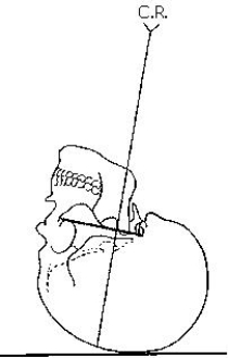
What is the central-ray angulation for demonstration of the entire foramen magnum during an AP axial (Towne) projection?
A) 37 degrees caudad
B) 40 degrees caudad
C) 60 degrees caudad
D) 40 to 60 degrees caudad
A) 37 degrees caudad
B) 40 degrees caudad
C) 60 degrees caudad
D) 40 to 60 degrees caudad
فتح الحزمة
افتح القفل للوصول البطاقات البالغ عددها 196 في هذه المجموعة.
فتح الحزمة
k this deck
43
For an SMV projection of the cranial base, the central ray should always be perpendicular to the _____ line.
A) mentomeatal
B) orbitomeatal
C) infraorbitomeatal
D) acanthiomeatal
A) mentomeatal
B) orbitomeatal
C) infraorbitomeatal
D) acanthiomeatal
فتح الحزمة
افتح القفل للوصول البطاقات البالغ عددها 196 في هذه المجموعة.
فتح الحزمة
k this deck
44
Which method of examining the skull is identified in the figure below?
A) Haas
B) Towne
C) Shüller
D) Caldwell
A) Haas
B) Towne
C) Shüller
D) Caldwell
فتح الحزمة
افتح القفل للوصول البطاقات البالغ عددها 196 في هذه المجموعة.
فتح الحزمة
k this deck
45
Which of the following is clearly demonstrated within the foramen magnum during an AP axial (Towne) projection of the skull?
1)Dorsum sellae
2)Sella turcica
3)Posterior clinoid processes
A) 1 and 2
B) 1 and 3
C) 2 and 3
D) 1, 2, and 3
1)Dorsum sellae
2)Sella turcica
3)Posterior clinoid processes
A) 1 and 2
B) 1 and 3
C) 2 and 3
D) 1, 2, and 3
فتح الحزمة
افتح القفل للوصول البطاقات البالغ عددها 196 في هذه المجموعة.
فتح الحزمة
k this deck
46
Which line should be placed parallel to the plane of the image receptor for the SMV projection of the cranial base?
A) Acanthiomeatal line
B) Orbitomeatal line
C) Infraorbitomeatal line
D) Mentomeatal line
A) Acanthiomeatal line
B) Orbitomeatal line
C) Infraorbitomeatal line
D) Mentomeatal line
فتح الحزمة
افتح القفل للوصول البطاقات البالغ عددها 196 في هذه المجموعة.
فتح الحزمة
k this deck
47
Radiographic demonstration of the cranial base is performed by which method?
A) Haas
B) Rhese
C) Towne
D) Schüller
A) Haas
B) Rhese
C) Towne
D) Schüller
فتح الحزمة
افتح القفل للوصول البطاقات البالغ عددها 196 في هذه المجموعة.
فتح الحزمة
k this deck
48
Which of the following is perpendicular to the image receptor plane for a Caldwell projection of the skull?
A) Glabellomeatal line
B) Acanthiomeatal line
C) Orbitomeatal line
D) Mentomeatal line
A) Glabellomeatal line
B) Acanthiomeatal line
C) Orbitomeatal line
D) Mentomeatal line
فتح الحزمة
افتح القفل للوصول البطاقات البالغ عددها 196 في هذه المجموعة.
فتح الحزمة
k this deck
49
What is the central-ray angulation for the SMV projection?
A) 0 degrees
B) 5 degrees caudad
C) 5 degrees cephalad
D) 5 to 7 degrees cephalad
A) 0 degrees
B) 5 degrees caudad
C) 5 degrees cephalad
D) 5 to 7 degrees cephalad
فتح الحزمة
افتح القفل للوصول البطاقات البالغ عددها 196 في هذه المجموعة.
فتح الحزمة
k this deck
50
If the patient cannot flex the neck to place the orbitomeatal line perpendicular to the image receptor for an AP axial (Towne) projection, which line should be placed perpendicular?
A) Acanthiomeatal line
B) Infraorbitomeatal line
C) Glabellomeatal line
D) Mentomeatal line
A) Acanthiomeatal line
B) Infraorbitomeatal line
C) Glabellomeatal line
D) Mentomeatal line
فتح الحزمة
افتح القفل للوصول البطاقات البالغ عددها 196 في هذه المجموعة.
فتح الحزمة
k this deck
51
If the infraorbitomeatal line is placed perpendicular to the image receptor during an AP axial (Towne) projection of the skull, how much is the central ray angled?
A) 15 degrees caudad
B) 30 degrees caudad
C) 37 degrees caudad
D) 45 degrees caudad
A) 15 degrees caudad
B) 30 degrees caudad
C) 37 degrees caudad
D) 45 degrees caudad
فتح الحزمة
افتح القفل للوصول البطاقات البالغ عددها 196 في هذه المجموعة.
فتح الحزمة
k this deck
52
The bones of the cranium are joined together by fibrous joints called:
A) diploë.
B) sulci.
C) sutures.
D) cartilage.
A) diploë.
B) sulci.
C) sutures.
D) cartilage.
فتح الحزمة
افتح القفل للوصول البطاقات البالغ عددها 196 في هذه المجموعة.
فتح الحزمة
k this deck
53
Often a patient cannot be turned into the prone position for a PA axial projection of the skull (Caldwell method). What central-ray angle would be used if the AP axial projection is used instead?
A) 10 degrees caudad
B) 15 degrees cephalad
C) 10 to 15 degrees caudad
D) 10 to 15 degrees cephalad
A) 10 degrees caudad
B) 15 degrees cephalad
C) 10 to 15 degrees caudad
D) 10 to 15 degrees cephalad
فتح الحزمة
افتح القفل للوصول البطاقات البالغ عددها 196 في هذه المجموعة.
فتح الحزمة
k this deck
54
What is the average central-ray angulation for the PA axial (Haas) projection of the skull?
A) 25 degrees caudad
B) 25 degrees cephalad
C) 30 degrees caudad
D) 30 degrees cephalad
A) 25 degrees caudad
B) 25 degrees cephalad
C) 30 degrees caudad
D) 30 degrees cephalad
فتح الحزمة
افتح القفل للوصول البطاقات البالغ عددها 196 في هذه المجموعة.
فتح الحزمة
k this deck
55
Which of the following is (are) clearly demonstrated on an SMV projection of the cranial base?
1)Mastoid process
2)Sphenoid process
3)Carotid canals
A) 1 and 2
B) 1 and 3
C) 2 and 3
D) 1, 2, and 3
1)Mastoid process
2)Sphenoid process
3)Carotid canals
A) 1 and 2
B) 1 and 3
C) 2 and 3
D) 1, 2, and 3
فتح الحزمة
افتح القفل للوصول البطاقات البالغ عددها 196 في هذه المجموعة.
فتح الحزمة
k this deck
56
Which of the following methods will clearly demonstrate the petrous ridges, foramen magnum, dorsum sellae, and posterior clinical processes?
1)Haas (PA axial)
2)Towne (AP axial)
3)Schüller (SMV)
A) 1 and 2
B) 1 and 3
C) 2 and 3
D) 1, 2, and 3
1)Haas (PA axial)
2)Towne (AP axial)
3)Schüller (SMV)
A) 1 and 2
B) 1 and 3
C) 2 and 3
D) 1, 2, and 3
فتح الحزمة
افتح القفل للوصول البطاقات البالغ عددها 196 في هذه المجموعة.
فتح الحزمة
k this deck
57
Which of the following is true regarding the placement of the image receptor for an AP axial (Towne) projection of the skull?
1)Its upper margin is at the level of the top of the cranium.
2)Its upper margin is 2 inches above the top of the cranium.
3)Its upper margin is 2 inches below the top of the cranium.
A) 1 only
B) 2 only
C) 3 only
D) 1, 2, and 3
1)Its upper margin is at the level of the top of the cranium.
2)Its upper margin is 2 inches above the top of the cranium.
3)Its upper margin is 2 inches below the top of the cranium.
A) 1 only
B) 2 only
C) 3 only
D) 1, 2, and 3
فتح الحزمة
افتح القفل للوصول البطاقات البالغ عددها 196 في هذه المجموعة.
فتح الحزمة
k this deck
58
Which of the following bones is contained in the floor of the cranium?
1)Ethmoid
2)Sphenoid
3)Temporal
A) 1 and 2
B) 1 and 3
C) 2 and 3
D) 1, 2, and 3
1)Ethmoid
2)Sphenoid
3)Temporal
A) 1 and 2
B) 1 and 3
C) 2 and 3
D) 1, 2, and 3
فتح الحزمة
افتح القفل للوصول البطاقات البالغ عددها 196 في هذه المجموعة.
فتح الحزمة
k this deck
59
Which of the following lines is placed perpendicular to the image receptor plane for the AP axial (Towne) projection?
A) Orbitomeatal line
B) Infraorbitomeatal line
C) Glabellomeatal line
D) Acanthiomeatal line
A) Orbitomeatal line
B) Infraorbitomeatal line
C) Glabellomeatal line
D) Acanthiomeatal line
فتح الحزمة
افتح القفل للوصول البطاقات البالغ عددها 196 في هذه المجموعة.
فتح الحزمة
k this deck
60
The x-ray projection demonstrated in the figure below is the:
A) SMV.
B) VSM.
C) AP axial.
D) PA axial.
A) SMV.
B) VSM.
C) AP axial.
D) PA axial.
فتح الحزمة
افتح القفل للوصول البطاقات البالغ عددها 196 في هذه المجموعة.
فتح الحزمة
k this deck
61
Which facial bone contains a foramen through which the tear duct passes?
A) Nasal
B) Palatine
C) Maxilla
D) Lacrimal
A) Nasal
B) Palatine
C) Maxilla
D) Lacrimal
فتح الحزمة
افتح القفل للوصول البطاقات البالغ عددها 196 في هذه المجموعة.
فتح الحزمة
k this deck
62
The bone identified on the skull below is the:
A) temporal.
B) parietal.
C) zygoma.
D) sphenoid.
A) temporal.
B) parietal.
C) zygoma.
D) sphenoid.
فتح الحزمة
افتح القفل للوصول البطاقات البالغ عددها 196 في هذه المجموعة.
فتح الحزمة
k this deck
63
The opening into the apex of the orbit for the transmission of the optic nerve and ophthalmic artery is called the:
A) optic canal.
B) foramen.
C) foramina ovale.
D) foramina rotundum.
A) optic canal.
B) foramen.
C) foramina ovale.
D) foramina rotundum.
فتح الحزمة
افتح القفل للوصول البطاقات البالغ عددها 196 في هذه المجموعة.
فتح الحزمة
k this deck
64
The part of the sphenoid bone identified in the figure below is the:
A) clivus.
B) foramen magnum.
C) sella turcica.
D) dorsum sellae.
A) clivus.
B) foramen magnum.
C) sella turcica.
D) dorsum sellae.
فتح الحزمة
افتح القفل للوصول البطاقات البالغ عددها 196 في هذه المجموعة.
فتح الحزمة
k this deck
65
Which of the following bones are contained in the calvarium?
1)Frontal
2)Parietal
3)Temporal
A) 1 and 2
B) 1 and 3
C) 2 and 3
D) 1, 2, and 3
1)Frontal
2)Parietal
3)Temporal
A) 1 and 2
B) 1 and 3
C) 2 and 3
D) 1, 2, and 3
فتح الحزمة
افتح القفل للوصول البطاقات البالغ عددها 196 في هذه المجموعة.
فتح الحزمة
k this deck
66
What type of joint is the TMJ?
A) Synovial-hinge
B) Synovial-gliding
C) Synovial-ellipsoidal
D) Synovial-hinge and gliding
A) Synovial-hinge
B) Synovial-gliding
C) Synovial-ellipsoidal
D) Synovial-hinge and gliding
فتح الحزمة
افتح القفل للوصول البطاقات البالغ عددها 196 في هذه المجموعة.
فتح الحزمة
k this deck
67
The large aperture in the occipital bone, through which the medulla oblongata and spinal cord exit, is termed the:
A) foramen magnum.
B) basilar part.
C) occipital protuberance.
D) hypoglossal canal.
A) foramen magnum.
B) basilar part.
C) occipital protuberance.
D) hypoglossal canal.
فتح الحزمة
افتح القفل للوصول البطاقات البالغ عددها 196 في هذه المجموعة.
فتح الحزمة
k this deck
68
The part of the sphenoid bone identified in the figure below is the:
A) clivus.
B) clinoid processes.
C) sella turcica.
D) dorsum sellae.
A) clivus.
B) clinoid processes.
C) sella turcica.
D) dorsum sellae.
فتح الحزمة
افتح القفل للوصول البطاقات البالغ عددها 196 في هذه المجموعة.
فتح الحزمة
k this deck
69
The suture located between the occipital bone and the parietal bones is the:
A) lambdoidal.
B) squamosal.
C) sagittal.
D) corona.
A) lambdoidal.
B) squamosal.
C) sagittal.
D) corona.
فتح الحزمة
افتح القفل للوصول البطاقات البالغ عددها 196 في هذه المجموعة.
فتح الحزمة
k this deck
70
The base of the anterior portion of the occipital bone contains two large openings that allow blood vessels and nerves to pass through. These two openings are called the:
A) jugular foramina.
B) foramen magnum.
C) foramen ovale.
D) hypoglossal canal.
A) jugular foramina.
B) foramen magnum.
C) foramen ovale.
D) hypoglossal canal.
فتح الحزمة
افتح القفل للوصول البطاقات البالغ عددها 196 في هذه المجموعة.
فتح الحزمة
k this deck
71
Which parts of the patient's face touch the table for a PA axial projection (Caldwell method)?
1)Forehead
2)Nose
3)Chin
A) 1 and 2
B) 1 and 3
C) 2 and 3
D) 1, 2, and 3
1)Forehead
2)Nose
3)Chin
A) 1 and 2
B) 1 and 3
C) 2 and 3
D) 1, 2, and 3
فتح الحزمة
افتح القفل للوصول البطاقات البالغ عددها 196 في هذه المجموعة.
فتح الحزمة
k this deck
72
The posterior half of the base of the skull is formed by which bone?
A) Temporal
B) Sphenoid
C) Occipital
D) Parietal
A) Temporal
B) Sphenoid
C) Occipital
D) Parietal
فتح الحزمة
افتح القفل للوصول البطاقات البالغ عددها 196 في هذه المجموعة.
فتح الحزمة
k this deck
73
Which two facial bones form the roof of the mouth?
1)Maxillae
2)Vomer
3)Palatine bones
A) 1 and 2
B) 1 and 3
C) 2 and 3
D) 1, 2, and 3
1)Maxillae
2)Vomer
3)Palatine bones
A) 1 and 2
B) 1 and 3
C) 2 and 3
D) 1, 2, and 3
فتح الحزمة
افتح القفل للوصول البطاقات البالغ عددها 196 في هذه المجموعة.
فتح الحزمة
k this deck
74
The thickest and densest portion of bone in the cranium is the _____ bone.
A) mastoid portion of the temporal
B) petrous portion of the temporal
C) basilar part of the occipital
D) glabella of the frontal
A) mastoid portion of the temporal
B) petrous portion of the temporal
C) basilar part of the occipital
D) glabella of the frontal
فتح الحزمة
افتح القفل للوصول البطاقات البالغ عددها 196 في هذه المجموعة.
فتح الحزمة
k this deck
75
Which of the following foramina lie in the sphenoid bone?
1)Optic foramen
2)Jugular foramen
3)Foramina rotundum
A) 1 and 2
B) 1 and 3
C) 2 and 3
D) 1, 2, and 3
1)Optic foramen
2)Jugular foramen
3)Foramina rotundum
A) 1 and 2
B) 1 and 3
C) 2 and 3
D) 1, 2, and 3
فتح الحزمة
افتح القفل للوصول البطاقات البالغ عددها 196 في هذه المجموعة.
فتح الحزمة
k this deck
76
The suture identified on the figure below is the:
A) coronal.
B) squamosal.
C) sagittal.
D) lambdoidal.
A) coronal.
B) squamosal.
C) sagittal.
D) lambdoidal.
فتح الحزمة
افتح القفل للوصول البطاقات البالغ عددها 196 في هذه المجموعة.
فتح الحزمة
k this deck
77
The superior aspect of the sphenoid bone contains a deep depression that contains the:
A) pituitary gland.
B) pineal gland.
C) carotid sulcus.
D) optic canal.
A) pituitary gland.
B) pineal gland.
C) carotid sulcus.
D) optic canal.
فتح الحزمة
افتح القفل للوصول البطاقات البالغ عددها 196 في هذه المجموعة.
فتح الحزمة
k this deck
78
The base of the temporal bone contains an opening through which the internal carotid artery passes and is termed the:
A) foramen spinosum.
B) foramen ovale.
C) foramen lacerum.
D) jugular foramen.
A) foramen spinosum.
B) foramen ovale.
C) foramen lacerum.
D) jugular foramen.
فتح الحزمة
افتح القفل للوصول البطاقات البالغ عددها 196 في هذه المجموعة.
فتح الحزمة
k this deck
79
The bone identified on the skull below is the:
A) temporal.
B) parietal.
C) occipital.
D) sphenoid.
A) temporal.
B) parietal.
C) occipital.
D) sphenoid.
فتح الحزمة
افتح القفل للوصول البطاقات البالغ عددها 196 في هذه المجموعة.
فتح الحزمة
k this deck
80
The six areas of incomplete ossification in a newborn infant's skull are called the:
A) sulci.
B) sutures.
C) diploë.
D) fontanels.
A) sulci.
B) sutures.
C) diploë.
D) fontanels.
فتح الحزمة
افتح القفل للوصول البطاقات البالغ عددها 196 في هذه المجموعة.
فتح الحزمة
k this deck
فتح الحزمة
افتح القفل للوصول البطاقات البالغ عددها 196 في هذه المجموعة.


