Deck 28: Sectional Anatomy for Radiographers
ملء الشاشة (f)
سؤال
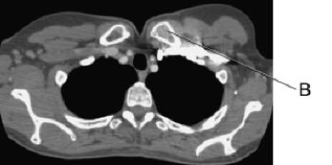
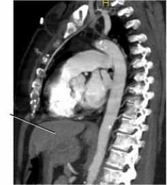
In this cross section of the thorax, line B identifies the:
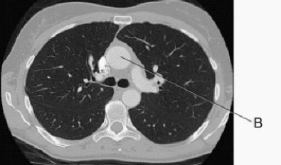
A) bronchus.
B) esophagus.
C) ascending aorta.
D) descending aorta.
A) bronchus.
B) esophagus.
C) ascending aorta.
D) descending aorta.
سؤال
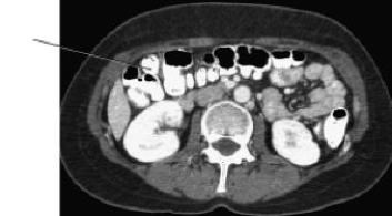
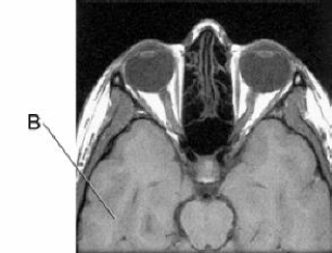
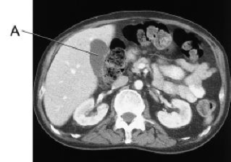
Line B on this image is pointing to the:
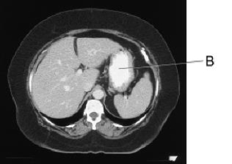
A) liver.
B) stomach.
C) spleen.
D) small bowel.
A) liver.
B) stomach.
C) spleen.
D) small bowel.
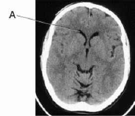
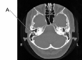
سؤال
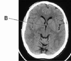
Line A is pointing to the:
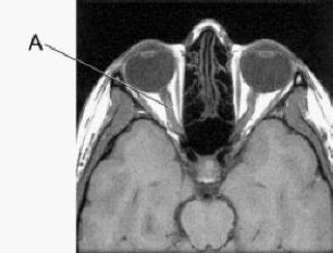
A) optic nerve.
B) pituitary gland.
C) carotid artery.
D) carotid vein.
A) optic nerve.
B) pituitary gland.
C) carotid artery.
D) carotid vein.
سؤال
This image is a(n) _____ plane of the _____ abdomen.
A) axial; upper
B) coronal; upper
C) axial; lower
D) coronal; lower
A) axial; upper
B) coronal; upper
C) axial; lower
D) coronal; lower
سؤال
Line B is pointing to the:
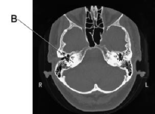
A) inner ear.
B) tympanic membrane.
C) internal auditory canal.
D) petrous portion of the temporal bone.
A) inner ear.
B) tympanic membrane.
C) internal auditory canal.
D) petrous portion of the temporal bone.
سؤال
Line B on this image is pointing to the:
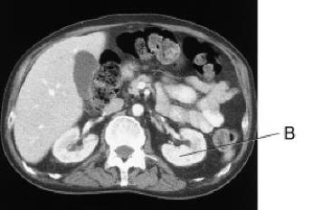
A) liver.
B) spleen.
C) L kidney.
D) gallbladder.
A) liver.
B) spleen.
C) L kidney.
D) gallbladder.
سؤال
Line A is pointing to the _____ in this CT image of the brain.
A) choroid plexus
B) third ventricle
C) lateral ventricle, anterior horn
D) lateral ventricle, occipital horn
A) choroid plexus
B) third ventricle
C) lateral ventricle, anterior horn
D) lateral ventricle, occipital horn
سؤال
This CT image of the brain is in which plane?
A) Axial
B) Coronal
C) Sagittal
D) Midsagittal
A) Axial
B) Coronal
C) Sagittal
D) Midsagittal
سؤال
Line B in this axial CT scan identifies the left:
A) clavicle.
B) brachiocephalic vein.
C) subclavian artery.
D) subclavian vein.
A) clavicle.
B) brachiocephalic vein.
C) subclavian artery.
D) subclavian vein.
سؤال
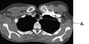
In this cross section of the thorax, line A identifies the:

A) bronchus.
B) esophagus.
C) ascending aorta.
D) descending aorta.
A) bronchus.
B) esophagus.
C) ascending aorta.
D) descending aorta.
سؤال
The line on this CT scan is pointing to the:
A) liver.
B) sternum.
C) stomach.
D) transverse colon.
A) liver.
B) sternum.
C) stomach.
D) transverse colon.
سؤال
Line A in this axial CT scan identifies the left:
A) clavicle.
B) brachiocephalic vein.
C) subclavian artery.
D) subclavian vein.
A) clavicle.
B) brachiocephalic vein.
C) subclavian artery.
D) subclavian vein.
سؤال
Line B is pointing to the _____ in this CT image of the brain.
A) pituitary gland
B) third ventricle
C) lateral ventricle
D) foramen of Monroe
A) pituitary gland
B) third ventricle
C) lateral ventricle
D) foramen of Monroe
سؤال
Line B is pointing to the:
A) pons.
B) cerebellum.
C) frontal lobe.
D) temporal lobe.
A) pons.
B) cerebellum.
C) frontal lobe.
D) temporal lobe.
سؤال
Line A on this image is pointing to the:

A) right lobe of the liver.
B) left lobe of the liver.
C) caudate lobe of the liver.
D) falciform ligament.
A) right lobe of the liver.
B) left lobe of the liver.
C) caudate lobe of the liver.
D) falciform ligament.
سؤال
The line on this CT scan of the facial bones identifies the _____ sinus.
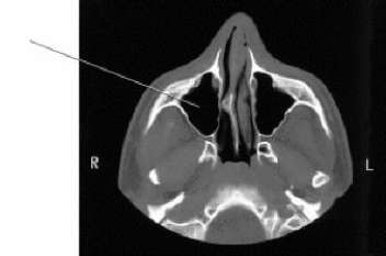
A) maxillary
B) sphenoid
C) middle ethmoid
D) posterior ethmoid
A) maxillary
B) sphenoid
C) middle ethmoid
D) posterior ethmoid
سؤال
The line on this image is pointing to the:

A) colon.
B) kidney.
C) stomach.
D) spleen.
A) colon.
B) kidney.
C) stomach.
D) spleen.
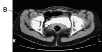
سؤال
Line A on this CT scan is pointing to the:
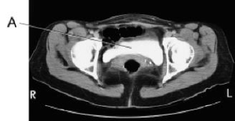
A) uterus.
B) rectum.
C) bladder.
D) small bowel.
A) uterus.
B) rectum.
C) bladder.
D) small bowel.
سؤال
Line A is pointing to the:
A) cochlea.
B) inner ear.
C) mastoid air cells.
D) internal auditory canal.
A) cochlea.
B) inner ear.
C) mastoid air cells.
D) internal auditory canal.
سؤال
Line A on this image is pointing to the:
A) liver.
B) spleen.
C) L kidney.
D) gallbladder.
A) liver.
B) spleen.
C) L kidney.
D) gallbladder.
سؤال
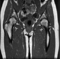
The line on this image of the pelvis identifies the:
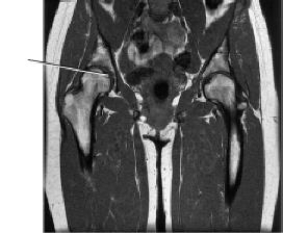
A) humerus.
B) glenoid cavity.
C) acetabulum.
D) greater trochanter.
A) humerus.
B) glenoid cavity.
C) acetabulum.
D) greater trochanter.
سؤال
The line on the image below points to the:
A) occipital lobe.
B) temporal lobe.
C) pons.
D) cerebellum.
A) occipital lobe.
B) temporal lobe.
C) pons.
D) cerebellum.
سؤال
Line B on this CT scan is pointing to the:
A) uterus.
B) rectum.
C) bladder.
D) small bowel.
A) uterus.
B) rectum.
C) bladder.
D) small bowel.
سؤال
The plane on this image is a(n):
A) coronal.
B) axial.
C) sagittal.
D) midcoronal.
A) coronal.
B) axial.
C) sagittal.
D) midcoronal.
سؤال
On the image below, the line points to the structure that forms the walls of the third ventricle, which is the:
A) pons.
B) thalamus.
C) lateral ventricle.
D) cerebellar peduncles.
A) pons.
B) thalamus.
C) lateral ventricle.
D) cerebellar peduncles.
سؤال
Examine the image below. The line points to the _____ muscle.
A) left psoas
B) right psoas
C) rectus abdominis
D) external oblique
A) left psoas
B) right psoas
C) rectus abdominis
D) external oblique
سؤال
Which of the following organs are demonstrated in the image below?
1)Spleen
2)Kidneys
3)Pancreas
A) 1 and 2
B) 1 and 3
C) 2 and 3
D) 1, 2, and 3
1)Spleen
2)Kidneys
3)Pancreas
A) 1 and 2
B) 1 and 3
C) 2 and 3
D) 1, 2, and 3
سؤال
Examine the image below. The line points to the:
A) stomach.
B) liver.
C) kidney.
D) heart.
A) stomach.
B) liver.
C) kidney.
D) heart.
سؤال
On the image below, the line points to the:
A) spinal cord.
B) ascending aorta.
C) descending aorta.
D) abdominal aorta.
A) spinal cord.
B) ascending aorta.
C) descending aorta.
D) abdominal aorta.
سؤال
The plane on this image is a(n):
A) coronal.
B) axial.
C) sagittal.
D) midsagittal.
A) coronal.
B) axial.
C) sagittal.
D) midsagittal.

فتح الحزمة
قم بالتسجيل لفتح البطاقات في هذه المجموعة!
Unlock Deck
Unlock Deck
1/30
العب
ملء الشاشة (f)
Deck 28: Sectional Anatomy for Radiographers
1
In this cross section of the thorax, line B identifies the:
A) bronchus.
B) esophagus.
C) ascending aorta.
D) descending aorta.
A) bronchus.
B) esophagus.
C) ascending aorta.
D) descending aorta.
ascending aorta.
2
Line B on this image is pointing to the:
A) liver.
B) stomach.
C) spleen.
D) small bowel.
A) liver.
B) stomach.
C) spleen.
D) small bowel.
stomach.
3
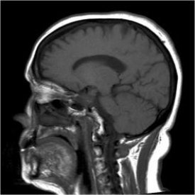
Line A is pointing to the:
A) optic nerve.
B) pituitary gland.
C) carotid artery.
D) carotid vein.
A) optic nerve.
B) pituitary gland.
C) carotid artery.
D) carotid vein.
optic nerve.
4
This image is a(n) _____ plane of the _____ abdomen.
A) axial; upper
B) coronal; upper
C) axial; lower
D) coronal; lower
A) axial; upper
B) coronal; upper
C) axial; lower
D) coronal; lower
فتح الحزمة
افتح القفل للوصول البطاقات البالغ عددها 30 في هذه المجموعة.
فتح الحزمة
k this deck
5
Line B is pointing to the:
A) inner ear.
B) tympanic membrane.
C) internal auditory canal.
D) petrous portion of the temporal bone.
A) inner ear.
B) tympanic membrane.
C) internal auditory canal.
D) petrous portion of the temporal bone.
فتح الحزمة
افتح القفل للوصول البطاقات البالغ عددها 30 في هذه المجموعة.
فتح الحزمة
k this deck
6
Line B on this image is pointing to the:
A) liver.
B) spleen.
C) L kidney.
D) gallbladder.
A) liver.
B) spleen.
C) L kidney.
D) gallbladder.
فتح الحزمة
افتح القفل للوصول البطاقات البالغ عددها 30 في هذه المجموعة.
فتح الحزمة
k this deck
7
Line A is pointing to the _____ in this CT image of the brain.
A) choroid plexus
B) third ventricle
C) lateral ventricle, anterior horn
D) lateral ventricle, occipital horn
A) choroid plexus
B) third ventricle
C) lateral ventricle, anterior horn
D) lateral ventricle, occipital horn
فتح الحزمة
افتح القفل للوصول البطاقات البالغ عددها 30 في هذه المجموعة.
فتح الحزمة
k this deck
8
This CT image of the brain is in which plane?
A) Axial
B) Coronal
C) Sagittal
D) Midsagittal
A) Axial
B) Coronal
C) Sagittal
D) Midsagittal
فتح الحزمة
افتح القفل للوصول البطاقات البالغ عددها 30 في هذه المجموعة.
فتح الحزمة
k this deck
9
Line B in this axial CT scan identifies the left:
A) clavicle.
B) brachiocephalic vein.
C) subclavian artery.
D) subclavian vein.
A) clavicle.
B) brachiocephalic vein.
C) subclavian artery.
D) subclavian vein.
فتح الحزمة
افتح القفل للوصول البطاقات البالغ عددها 30 في هذه المجموعة.
فتح الحزمة
k this deck
10
In this cross section of the thorax, line A identifies the:
A) bronchus.
B) esophagus.
C) ascending aorta.
D) descending aorta.
A) bronchus.
B) esophagus.
C) ascending aorta.
D) descending aorta.
فتح الحزمة
افتح القفل للوصول البطاقات البالغ عددها 30 في هذه المجموعة.
فتح الحزمة
k this deck
11
The line on this CT scan is pointing to the:
A) liver.
B) sternum.
C) stomach.
D) transverse colon.
A) liver.
B) sternum.
C) stomach.
D) transverse colon.
فتح الحزمة
افتح القفل للوصول البطاقات البالغ عددها 30 في هذه المجموعة.
فتح الحزمة
k this deck
12
Line A in this axial CT scan identifies the left:
A) clavicle.
B) brachiocephalic vein.
C) subclavian artery.
D) subclavian vein.
A) clavicle.
B) brachiocephalic vein.
C) subclavian artery.
D) subclavian vein.
فتح الحزمة
افتح القفل للوصول البطاقات البالغ عددها 30 في هذه المجموعة.
فتح الحزمة
k this deck
13
Line B is pointing to the _____ in this CT image of the brain.
A) pituitary gland
B) third ventricle
C) lateral ventricle
D) foramen of Monroe
A) pituitary gland
B) third ventricle
C) lateral ventricle
D) foramen of Monroe
فتح الحزمة
افتح القفل للوصول البطاقات البالغ عددها 30 في هذه المجموعة.
فتح الحزمة
k this deck
14
Line B is pointing to the:
A) pons.
B) cerebellum.
C) frontal lobe.
D) temporal lobe.
A) pons.
B) cerebellum.
C) frontal lobe.
D) temporal lobe.
فتح الحزمة
افتح القفل للوصول البطاقات البالغ عددها 30 في هذه المجموعة.
فتح الحزمة
k this deck
15
Line A on this image is pointing to the:
A) right lobe of the liver.
B) left lobe of the liver.
C) caudate lobe of the liver.
D) falciform ligament.
A) right lobe of the liver.
B) left lobe of the liver.
C) caudate lobe of the liver.
D) falciform ligament.
فتح الحزمة
افتح القفل للوصول البطاقات البالغ عددها 30 في هذه المجموعة.
فتح الحزمة
k this deck
16
The line on this CT scan of the facial bones identifies the _____ sinus.
A) maxillary
B) sphenoid
C) middle ethmoid
D) posterior ethmoid
A) maxillary
B) sphenoid
C) middle ethmoid
D) posterior ethmoid
فتح الحزمة
افتح القفل للوصول البطاقات البالغ عددها 30 في هذه المجموعة.
فتح الحزمة
k this deck
17
The line on this image is pointing to the:
A) colon.
B) kidney.
C) stomach.
D) spleen.
A) colon.
B) kidney.
C) stomach.
D) spleen.
فتح الحزمة
افتح القفل للوصول البطاقات البالغ عددها 30 في هذه المجموعة.
فتح الحزمة
k this deck
18
Line A on this CT scan is pointing to the:
A) uterus.
B) rectum.
C) bladder.
D) small bowel.
A) uterus.
B) rectum.
C) bladder.
D) small bowel.
فتح الحزمة
افتح القفل للوصول البطاقات البالغ عددها 30 في هذه المجموعة.
فتح الحزمة
k this deck
19
Line A is pointing to the:
A) cochlea.
B) inner ear.
C) mastoid air cells.
D) internal auditory canal.
A) cochlea.
B) inner ear.
C) mastoid air cells.
D) internal auditory canal.
فتح الحزمة
افتح القفل للوصول البطاقات البالغ عددها 30 في هذه المجموعة.
فتح الحزمة
k this deck
20
Line A on this image is pointing to the:
A) liver.
B) spleen.
C) L kidney.
D) gallbladder.
A) liver.
B) spleen.
C) L kidney.
D) gallbladder.
فتح الحزمة
افتح القفل للوصول البطاقات البالغ عددها 30 في هذه المجموعة.
فتح الحزمة
k this deck
21
The line on this image of the pelvis identifies the:
A) humerus.
B) glenoid cavity.
C) acetabulum.
D) greater trochanter.
A) humerus.
B) glenoid cavity.
C) acetabulum.
D) greater trochanter.
فتح الحزمة
افتح القفل للوصول البطاقات البالغ عددها 30 في هذه المجموعة.
فتح الحزمة
k this deck
22
The line on the image below points to the:
A) occipital lobe.
B) temporal lobe.
C) pons.
D) cerebellum.
A) occipital lobe.
B) temporal lobe.
C) pons.
D) cerebellum.
فتح الحزمة
افتح القفل للوصول البطاقات البالغ عددها 30 في هذه المجموعة.
فتح الحزمة
k this deck
23
Line B on this CT scan is pointing to the:
A) uterus.
B) rectum.
C) bladder.
D) small bowel.
A) uterus.
B) rectum.
C) bladder.
D) small bowel.
فتح الحزمة
افتح القفل للوصول البطاقات البالغ عددها 30 في هذه المجموعة.
فتح الحزمة
k this deck
24
The plane on this image is a(n):
A) coronal.
B) axial.
C) sagittal.
D) midcoronal.
A) coronal.
B) axial.
C) sagittal.
D) midcoronal.
فتح الحزمة
افتح القفل للوصول البطاقات البالغ عددها 30 في هذه المجموعة.
فتح الحزمة
k this deck
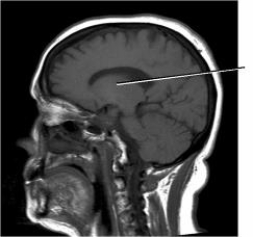
25
On the image below, the line points to the structure that forms the walls of the third ventricle, which is the:
A) pons.
B) thalamus.
C) lateral ventricle.
D) cerebellar peduncles.
A) pons.
B) thalamus.
C) lateral ventricle.
D) cerebellar peduncles.
فتح الحزمة
افتح القفل للوصول البطاقات البالغ عددها 30 في هذه المجموعة.
فتح الحزمة
k this deck
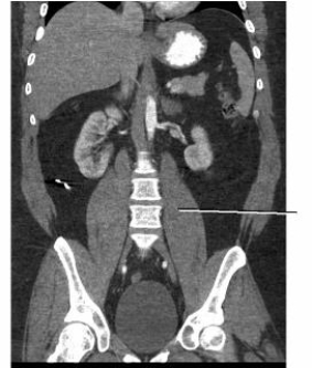
26
Examine the image below. The line points to the _____ muscle.
A) left psoas
B) right psoas
C) rectus abdominis
D) external oblique
A) left psoas
B) right psoas
C) rectus abdominis
D) external oblique
فتح الحزمة
افتح القفل للوصول البطاقات البالغ عددها 30 في هذه المجموعة.
فتح الحزمة
k this deck
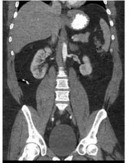
27
Which of the following organs are demonstrated in the image below?
1)Spleen
2)Kidneys
3)Pancreas
A) 1 and 2
B) 1 and 3
C) 2 and 3
D) 1, 2, and 3
1)Spleen
2)Kidneys
3)Pancreas
A) 1 and 2
B) 1 and 3
C) 2 and 3
D) 1, 2, and 3
فتح الحزمة
افتح القفل للوصول البطاقات البالغ عددها 30 في هذه المجموعة.
فتح الحزمة
k this deck
28
Examine the image below. The line points to the:
A) stomach.
B) liver.
C) kidney.
D) heart.
A) stomach.
B) liver.
C) kidney.
D) heart.
فتح الحزمة
افتح القفل للوصول البطاقات البالغ عددها 30 في هذه المجموعة.
فتح الحزمة
k this deck
29
On the image below, the line points to the:
A) spinal cord.
B) ascending aorta.
C) descending aorta.
D) abdominal aorta.
A) spinal cord.
B) ascending aorta.
C) descending aorta.
D) abdominal aorta.
فتح الحزمة
افتح القفل للوصول البطاقات البالغ عددها 30 في هذه المجموعة.
فتح الحزمة
k this deck
30
The plane on this image is a(n):
A) coronal.
B) axial.
C) sagittal.
D) midsagittal.
A) coronal.
B) axial.
C) sagittal.
D) midsagittal.
فتح الحزمة
افتح القفل للوصول البطاقات البالغ عددها 30 في هذه المجموعة.
فتح الحزمة
k this deck
فتح الحزمة
افتح القفل للوصول البطاقات البالغ عددها 30 في هذه المجموعة.


