Deck 27: Chest Patterns
ملء الشاشة (f)
سؤال
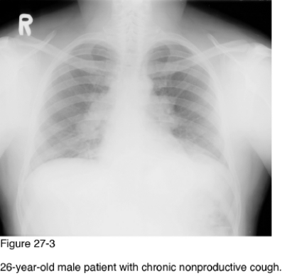
The appearance of the hilar regions in Figure 27-3 is best described as __________.
A) Batwing
B) Potato nodes
C) Kerley C lines
D) Ranke complex
سؤال
Which radiographic sign accounts for part non-visualization of the right heart border on the PA radiograph?
A) S-shaped Golden
B) Luftsichel
C) Plate
D) Silhouette
سؤال
Which of the following conditions would be most likely if enlarged right paratracheal nodes were also present on the radiographs and Figure 27-3?
A) Bronchogenic carcinoma
B) Lymphoma
C) Leukemia
D) Sarcoidosis
سؤال
What radiographic pattern of disease is present in Figure 27-3?
A) Localized airspace disease
B) Enlarged hilum
C) Chest wall and pleural-based lesions
D) Multiple nodules and masses
سؤال
On the basis of the position of the lesion in the periphery of the lung parenchyma, which of the following cell types is likely to make up the mass if the underlying cause is bronchogenic carcinoma?
A) Squamous cell
B) Adenocarcinoma
C) Alveolar cell carcinoma
D) Small cell carcinoma
سؤال
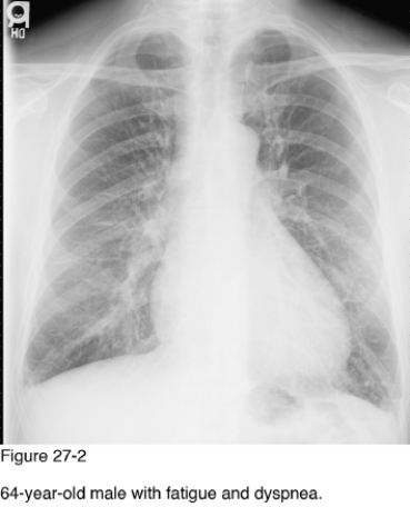
Which of the following best characterizes the radiographic pattern of lung disease and Figure 27-2?
A) Multiple nodules and masses
B) Diffuse alveolar disease
C) Enlarged hilum
D) Diffuse interstitial disease
سؤال
Which of the following diagnoses best accounts for the radiographic abnormalities in Figure 27-2?
A) Cardiogenic pulmonary edema
B) Aspiration pneumonia
C) Adult respiratory distress syndrome
D) Neurogenic pulmonary edema
سؤال
سؤال
The linear densities noted at the lateral margins of the lower lungs are known as __________.
A) Kerley A lines
B) Kerley B lines
C) Kerley C lines
D) Subsegmental atelectasis
سؤال
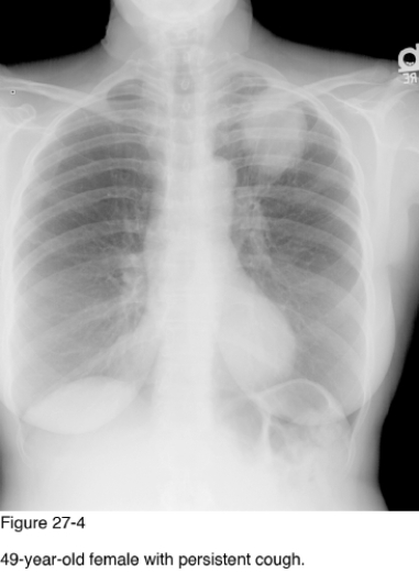
What radiographic pattern of disease is evident on Figure 27-4?
A) Multiple nodules and masses
B) Lung parenchymal calcifications
C) Solitary pulmonary nodule/mass
D) Pleural calcifications
سؤال
Which of the following diagnoses would be most likely if this patient disclosed a history of cigarette smoking?
A) Bronchiocarcinoid tumor
B) Bronchogenic carcinoma
C) Bronchogenic cyst
D) Metastasis
سؤال
Definitive diagnosis of the underlying cause of this patient's disease would best be accomplished with __________.
A) Biopsy
B) Computed tomography of the chest
C) Sputum culture
D) Spirometry
سؤال
On PA chest radiographs, what is a normal measure of the pulmonary arteries?
A) 5 mm
B) 11 mm
C) 16 mm
D) 22 mm
سؤال
Considering the patient's history, what is likely to be the underlying cause of this abnormality?
A) Lymphadenopathy
B) Mucous plug obstructing a bronchus
C) Air-space consolidation
D) Cardiomegaly
سؤال
What other clinical symptoms are likely in this patient?
A) Night sweats
B) Fever
C) Wheezing and dyspnea
D) No symptoms (asymptomatic)
سؤال
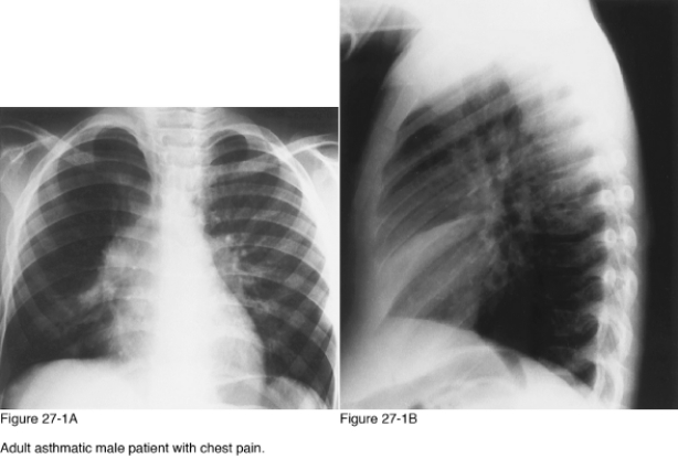
Which radiographic pattern of disease is evident in Figure 27-1?
A) Atelectasis
B) Hilar and mediastinal calcification
C) Enlarged hilum
D) Localized alveolar (air-space) disease
سؤال
Failure of which organ(s) may produce similar radiographic findings to those in Figure 27-2?
A) Kidneys
B) Splay
C) Pancreas
D) Liver
سؤال
Normal transverse dimension of the heart should measure less than __________ the widest transverse dimension of the thorax measured along the inner rib margins.
A) One fourth
B) One third
C) One half
D) Two thirds
سؤال
The triangular-shaped density noted on the lateral chest radiograph represents the right __________.
A) Hilum
B) Upper lobe
C) Middle lobe
D) Lower lobe
سؤال
Doubling of the lesion's size in which of the following timeframes would support a diagnosis of bronchogenic carcinoma?
A) 0-1 month
B) 1-18 months
C) 18-24 months
D) 24-36 months

فتح الحزمة
قم بالتسجيل لفتح البطاقات في هذه المجموعة!
Unlock Deck
Unlock Deck
1/20
العب
ملء الشاشة (f)
Deck 27: Chest Patterns
1
The appearance of the hilar regions in Figure 27-3 is best described as __________.
A) Batwing
B) Potato nodes
C) Kerley C lines
D) Ranke complex
Potato nodes
2
Which radiographic sign accounts for part non-visualization of the right heart border on the PA radiograph?
A) S-shaped Golden
B) Luftsichel
C) Plate
D) Silhouette
Silhouette
3
Which of the following conditions would be most likely if enlarged right paratracheal nodes were also present on the radiographs and Figure 27-3?
A) Bronchogenic carcinoma
B) Lymphoma
C) Leukemia
D) Sarcoidosis
Sarcoidosis
4
What radiographic pattern of disease is present in Figure 27-3?
A) Localized airspace disease
B) Enlarged hilum
C) Chest wall and pleural-based lesions
D) Multiple nodules and masses
فتح الحزمة
افتح القفل للوصول البطاقات البالغ عددها 20 في هذه المجموعة.
فتح الحزمة
k this deck
5
On the basis of the position of the lesion in the periphery of the lung parenchyma, which of the following cell types is likely to make up the mass if the underlying cause is bronchogenic carcinoma?
A) Squamous cell
B) Adenocarcinoma
C) Alveolar cell carcinoma
D) Small cell carcinoma
فتح الحزمة
افتح القفل للوصول البطاقات البالغ عددها 20 في هذه المجموعة.
فتح الحزمة
k this deck
6
Which of the following best characterizes the radiographic pattern of lung disease and Figure 27-2?
A) Multiple nodules and masses
B) Diffuse alveolar disease
C) Enlarged hilum
D) Diffuse interstitial disease
فتح الحزمة
افتح القفل للوصول البطاقات البالغ عددها 20 في هذه المجموعة.
فتح الحزمة
k this deck
7
Which of the following diagnoses best accounts for the radiographic abnormalities in Figure 27-2?
A) Cardiogenic pulmonary edema
B) Aspiration pneumonia
C) Adult respiratory distress syndrome
D) Neurogenic pulmonary edema
فتح الحزمة
افتح القفل للوصول البطاقات البالغ عددها 20 في هذه المجموعة.
فتح الحزمة
k this deck
8
Which of the following diagnostic imaging modalities would provide greatest sensitivity in further characterization of the lesion?
A) Computed tomography
B) Magnetic resonance imaging
C) Diagnostic ultrasound
D) Apical lordotic and lateral chest radiographs
A) Computed tomography
B) Magnetic resonance imaging
C) Diagnostic ultrasound
D) Apical lordotic and lateral chest radiographs
فتح الحزمة
افتح القفل للوصول البطاقات البالغ عددها 20 في هذه المجموعة.
فتح الحزمة
k this deck
9
The linear densities noted at the lateral margins of the lower lungs are known as __________.
A) Kerley A lines
B) Kerley B lines
C) Kerley C lines
D) Subsegmental atelectasis
فتح الحزمة
افتح القفل للوصول البطاقات البالغ عددها 20 في هذه المجموعة.
فتح الحزمة
k this deck
10
What radiographic pattern of disease is evident on Figure 27-4?
A) Multiple nodules and masses
B) Lung parenchymal calcifications
C) Solitary pulmonary nodule/mass
D) Pleural calcifications
فتح الحزمة
افتح القفل للوصول البطاقات البالغ عددها 20 في هذه المجموعة.
فتح الحزمة
k this deck
11
Which of the following diagnoses would be most likely if this patient disclosed a history of cigarette smoking?
A) Bronchiocarcinoid tumor
B) Bronchogenic carcinoma
C) Bronchogenic cyst
D) Metastasis
فتح الحزمة
افتح القفل للوصول البطاقات البالغ عددها 20 في هذه المجموعة.
فتح الحزمة
k this deck
12
Definitive diagnosis of the underlying cause of this patient's disease would best be accomplished with __________.
A) Biopsy
B) Computed tomography of the chest
C) Sputum culture
D) Spirometry
فتح الحزمة
افتح القفل للوصول البطاقات البالغ عددها 20 في هذه المجموعة.
فتح الحزمة
k this deck
13
On PA chest radiographs, what is a normal measure of the pulmonary arteries?
A) 5 mm
B) 11 mm
C) 16 mm
D) 22 mm
فتح الحزمة
افتح القفل للوصول البطاقات البالغ عددها 20 في هذه المجموعة.
فتح الحزمة
k this deck
14
Considering the patient's history, what is likely to be the underlying cause of this abnormality?
A) Lymphadenopathy
B) Mucous plug obstructing a bronchus
C) Air-space consolidation
D) Cardiomegaly
فتح الحزمة
افتح القفل للوصول البطاقات البالغ عددها 20 في هذه المجموعة.
فتح الحزمة
k this deck
15
What other clinical symptoms are likely in this patient?
A) Night sweats
B) Fever
C) Wheezing and dyspnea
D) No symptoms (asymptomatic)
فتح الحزمة
افتح القفل للوصول البطاقات البالغ عددها 20 في هذه المجموعة.
فتح الحزمة
k this deck
16
Which radiographic pattern of disease is evident in Figure 27-1?
A) Atelectasis
B) Hilar and mediastinal calcification
C) Enlarged hilum
D) Localized alveolar (air-space) disease
فتح الحزمة
افتح القفل للوصول البطاقات البالغ عددها 20 في هذه المجموعة.
فتح الحزمة
k this deck
17
Failure of which organ(s) may produce similar radiographic findings to those in Figure 27-2?
A) Kidneys
B) Splay
C) Pancreas
D) Liver
فتح الحزمة
افتح القفل للوصول البطاقات البالغ عددها 20 في هذه المجموعة.
فتح الحزمة
k this deck
18
Normal transverse dimension of the heart should measure less than __________ the widest transverse dimension of the thorax measured along the inner rib margins.
A) One fourth
B) One third
C) One half
D) Two thirds
فتح الحزمة
افتح القفل للوصول البطاقات البالغ عددها 20 في هذه المجموعة.
فتح الحزمة
k this deck
19
The triangular-shaped density noted on the lateral chest radiograph represents the right __________.
A) Hilum
B) Upper lobe
C) Middle lobe
D) Lower lobe
فتح الحزمة
افتح القفل للوصول البطاقات البالغ عددها 20 في هذه المجموعة.
فتح الحزمة
k this deck
20
Doubling of the lesion's size in which of the following timeframes would support a diagnosis of bronchogenic carcinoma?
A) 0-1 month
B) 1-18 months
C) 18-24 months
D) 24-36 months
فتح الحزمة
افتح القفل للوصول البطاقات البالغ عددها 20 في هذه المجموعة.
فتح الحزمة
k this deck
فتح الحزمة
افتح القفل للوصول البطاقات البالغ عددها 20 في هذه المجموعة.


