A 27-year-old woman is brought to the emergency department due to a 4-hour history of chest pain that is localized to the middle of the chest and the upper sternal area. She describes it as "intense." The patient has never before experienced a similar pain. She also describes some nausea and a mild occipital headache. She has had no vomiting, abdominal pain, or shortness of breath. A friend who accompanies her to the emergency department says that they attended a party and the patient smoked crack cocaine shortly prior to her episode of chest pain. She did not consume alcohol. The patient has a history of intravenous drug use. She was treated with antibiotics for upper extremity cellulitis 6 months ago. There is no family history of premature coronary artery disease. The patient does not take any medications and has no known drug allergies. On initial evaluation, temperature is 37.8 C (100 F) , blood pressure is 204/102 mm Hg on the right arm and 210/104 mm Hg on the left arm, pulse is 102/min and regular, and respirations are 18/min. Oxygen saturation is 99% on room air. The patient appears thin, anxious, and agitated. Heart sounds are normal and no murmurs are heard. Lungs are clear to auscultation bilaterally. The abdomen is soft and nontender. Lower extremity pulses are full and symmetric. There is no peripheral edema. ECG shows sinus tachycardia but is otherwise unremarkable. Portable chest x-ray reveals clear lung fields. Finger-stick blood glucose level is 98 mg/dL. After 2 hours in the emergency department, the patient has weakness in her right arm. Her chest pain is somewhat better, but she still has pain at the middle and upper part of the sternum. She has been treated with lorazepam, morphine, and nitroglycerin infusion. Blood pressure is 133/80 mm Hg and heart rate is 88/min and regular. Neurologic examination shows mild muscle weakness affecting the right upper and lower extremities. Speech is normal. Repeat ECG shows normal sinus rhythm with no significant abnormalities. Laboratory results are as follows: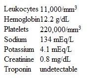 Immediate noncontrast CT scan of the head reveals no evidence of bleeding. Which of the following is the best next step in management of this patient?
A) Carotid artery ultrasound
B) CT angiography of the chest
C) Intravenous alteplase
D) Intravenous phentolamine
E) Low-molecular-weight heparin
Correct Answer:
Verified
Q97: A 62-year-old man is brought to the
Q98: A 70-year-old man comes to the hospital
Q99: A 56-year-old man is brought to the
Q100: A 57-year-old woman undergoes aortic valve replacement
Q101: A 52-year-old man comes to the emergency
Q103: A 72-year-old man comes to a rural
Q104: A 46-year-old woman comes to the clinic
Q105: A 50-year-old man is brought to the
Q106: A 42-year-old man comes to the office
Q107: A 68-year-old woman is referred for perioperative
Unlock this Answer For Free Now!
View this answer and more for free by performing one of the following actions

Scan the QR code to install the App and get 2 free unlocks

Unlock quizzes for free by uploading documents