Deck 14: The Nervous System: The Spinal Cord and Spinal Nerves
Full screen (f)
Question
Question
Question
Question
Question
Question
Question
Question
Question
Question
Question
Question
Question
Question
Question
Question
Question
Question
Question
Question
Question
Question
Question
Question
Question
Question
Question
Question
Question
Question
Question
Question
Question
Question
Question
Question
Question
Question
Question
Question
Question
Question
Question
Question
Question
Question
Question
Question
Question
Question
Question
Question
Question
Question
Question
Question
Question
Question
Question
Question
Question
Figure 14.2
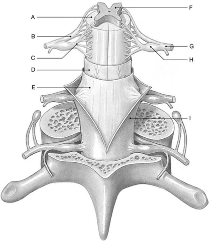
Using the above-referenced diagram of a posterior view of the spinal cord showing the meningeal layers, superficial landmarks, and distribution of gray and white matter, identify the specified labeled structure(s) in each of the following questions.
Identify the structure(s) indicated by Label A.
A) White matter
B) Dorsal horns
C) Dura mater
D) Spinal nerve
E) Ventral root
Question
Figure 14.2
Using the above-referenced diagram of a posterior view of the spinal cord showing the meningeal layers, superficial landmarks, and distribution of gray and white matter, identify the specified labeled structure(s) in each of the following questions.
Identify the structure(s) indicated by Label G.
A) Arachnoid mater
B) Dorsal root
C) Spinal nerve
D) Denticulate ligament
E) Ventral root
Question
Question
Question
Figure 14.2
Using the above-referenced diagram of a posterior view of the spinal cord showing the meningeal layers, superficial landmarks, and distribution of gray and white matter, identify the specified labeled structure(s) in each of the following questions.
Identify the structure(s) indicated by Label D.
A) Dura mater
B) Dorsal root
C) Arachnoid mater
D) Ventral root
E) Pia mater
Question
Figure 14.1
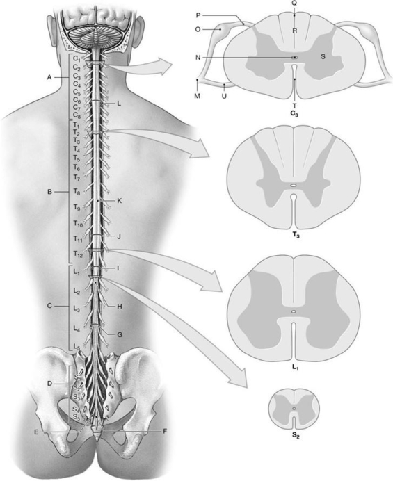
Using the above- referenced diagrams of the superficial anatomy and orientation of the adult spinal cord (posterior view)and inferior views of cross-sections through representative segments of the spinal cord, showing the arrangement of gray and white matter, identify the specified labeled structure(s) in each of the following questions.
Identify the structure(s) indicated by Label E.
A) Cervical spinal nerve
B) Coccygeal nerve
C) Thoracic spinal nerve
D) Lumbar spinal nerve
E) Inferior tip of the spinal cord
Question
Question
Figure 14.1
Using the above- referenced diagrams of the superficial anatomy and orientation of the adult spinal cord (posterior view)and inferior views of cross-sections through representative segments of the spinal cord, showing the arrangement of gray and white matter, identify the specified labeled structure(s) in each of the following questions.
Identify the structure(s) indicated by Label T.
A) Filum terminale
B) Anterior median fissure
C) Cauda equina
D) Conus medullaris
E) Posterior median sulcus
Question
Figure 14.1
Using the above- referenced diagrams of the superficial anatomy and orientation of the adult spinal cord (posterior view)and inferior views of cross-sections through representative segments of the spinal cord, showing the arrangement of gray and white matter, identify the specified labeled structure(s) in each of the following questions.
Identify the structure(s) indicated by Label N.
A) Central canal
B) Filum terminale
C) Cauda equina
D) Conus medullaris
E) Cervical spinal nerve
Question
Figure 14.1
Using the above- referenced diagrams of the superficial anatomy and orientation of the adult spinal cord (posterior view)and inferior views of cross-sections through representative segments of the spinal cord, showing the arrangement of gray and white matter, identify the specified labeled structure(s) in each of the following questions.
Identify the structure(s) indicated by Label G.
A) Coccygeal nerve
B) Lumbosacral enlargement
C) Filum terminale
D) Anterior median sulcus
E) Cauda equina
Question
Figure 14.2
Using the above-referenced diagram of a posterior view of the spinal cord showing the meningeal layers, superficial landmarks, and distribution of gray and white matter, identify the specified labeled structure(s) in each of the following questions.
Identify the structure(s) indicated by Label C.
A) Gray matter
B) Dorsal root
C) White matter
D) Ventral root
E) Spinal nerve
Question
Figure 14.2
Using the above-referenced diagram of a posterior view of the spinal cord showing the meningeal layers, superficial landmarks, and distribution of gray and white matter, identify the specified labeled structure(s) in each of the following questions.
Identify the structure(s) indicated by Label B.
A) Dorsal root
B) Denticulate ligament
C) Arachnoid mater
D) Ventral root
E) Dura mater
Question
Figure 14.2
Using the above-referenced diagram of a posterior view of the spinal cord showing the meningeal layers, superficial landmarks, and distribution of gray and white matter, identify the specified labeled structure(s) in each of the following questions.
Identify the structure(s) indicated by Label F.
A) Dorsal root
B) Ventral root
C) Dura mater
D) Gray matter
E) White matter
Question
Question
Question
Figure 14.1
Using the above- referenced diagrams of the superficial anatomy and orientation of the adult spinal cord (posterior view)and inferior views of cross-sections through representative segments of the spinal cord, showing the arrangement of gray and white matter, identify the specified labeled structure(s) in each of the following questions.
Identify the structure(s) indicated by Label K.
A) Anterior median sulcus
B) Cervical enlargement
C) Lumbosacral enlargement
D) Posterior median sulcus
E) Conus medullaris
Question
Figure 14.2
Using the above-referenced diagram of a posterior view of the spinal cord showing the meningeal layers, superficial landmarks, and distribution of gray and white matter, identify the specified labeled structure(s) in each of the following questions.
Identify the structure(s) indicated by Label E.
A) Pia mater
B) Arachnoid mater
C) Ventral root
D) Dura mater
E) Dorsal root
Question
Figure 14.1
Using the above- referenced diagrams of the superficial anatomy and orientation of the adult spinal cord (posterior view)and inferior views of cross-sections through representative segments of the spinal cord, showing the arrangement of gray and white matter, identify the specified labeled structure(s) in each of the following questions.
Identify the structure(s) indicated by Label I.
A) Cauda equina
B) Filum terminale
C) Lumbosacral enlargement
D) Conus medullaris
E) Coccygeal nerve
Question
Figure 14.1
Using the above- referenced diagrams of the superficial anatomy and orientation of the adult spinal cord (posterior view)and inferior views of cross-sections through representative segments of the spinal cord, showing the arrangement of gray and white matter, identify the specified labeled structure(s) in each of the following questions.
Identify the structure(s) indicated by Label F.
A) Filum terminale
B) Coccygeal nerve
C) Cervical spinal nerve
D) Posterior median sulcus
E) Lumbar spinal nerve
Question
Figure 14.1
Using the above- referenced diagrams of the superficial anatomy and orientation of the adult spinal cord (posterior view)and inferior views of cross-sections through representative segments of the spinal cord, showing the arrangement of gray and white matter, identify the specified labeled structure(s) in each of the following questions.
Identify the structure(s) indicated by Label O.
A) Ventral root
B) Dorsal root ganglion
C) Central canal
D) White matter
E) Anterior median fissure

Unlock Deck
Sign up to unlock the cards in this deck!
Unlock Deck
Unlock Deck
1/131
Play
Full screen (f)
Deck 14: The Nervous System: The Spinal Cord and Spinal Nerves
1
The epidural space contains areolar and adipose tissue, in addition to ________.
A) cerebrospinal fluid
B) lymph
C) denticulate ligaments
D) gray matter
E) blood vessels
A) cerebrospinal fluid
B) lymph
C) denticulate ligaments
D) gray matter
E) blood vessels
E
2
Inferior to the ________, the spinal cord tapers to the conus medullaris.
A) cervical enlargement
B) cauda equina
C) lumbosacral enlargement
D) filum terminale
E) dorsal root ganglia
A) cervical enlargement
B) cauda equina
C) lumbosacral enlargement
D) filum terminale
E) dorsal root ganglia
C
3
The pia mater is the ________.
A) meninx that forms denticulate ligaments
B) outermost covering over the brain, but not the spinal cord
C) toughest and thickest of the meninges
D) meninx that contains cerebrospinal fluid
E) meninx that forms the major component of the coccygeal ligament
A) meninx that forms denticulate ligaments
B) outermost covering over the brain, but not the spinal cord
C) toughest and thickest of the meninges
D) meninx that contains cerebrospinal fluid
E) meninx that forms the major component of the coccygeal ligament
A
4
In gross dissection, the filum terminale and the long ventral and dorsal roots are collectively referred to as the ________.
A) collateral ganglia
B) denticulate ligaments
C) cauda equina
D) spinal meninges
E) motor neurons
A) collateral ganglia
B) denticulate ligaments
C) cauda equina
D) spinal meninges
E) motor neurons
Unlock Deck
Unlock for access to all 131 flashcards in this deck.
Unlock Deck
k this deck
5
Caudally, the spinal dura mater blends with components of the ________ to form the coccygeal ligament.
A) dorsal root ganglia
B) filum terminale
C) ventral root
D) spinal nerves
E) cauda equina
A) dorsal root ganglia
B) filum terminale
C) ventral root
D) spinal nerves
E) cauda equina
Unlock Deck
Unlock for access to all 131 flashcards in this deck.
Unlock Deck
k this deck
6
The gray matter of the spinal cord is organized into anterior, posterior, and lateral ________.
A) tracts
B) columns
C) commissures
D) horns
E) ganglia
A) tracts
B) columns
C) commissures
D) horns
E) ganglia
Unlock Deck
Unlock for access to all 131 flashcards in this deck.
Unlock Deck
k this deck
7
Motor fibers travel in the ________ of the spinal cord.
A) ascending tracts
B) medial tracts
C) descending tracts
D) posterior tracts
E) lateral tracts
A) ascending tracts
B) medial tracts
C) descending tracts
D) posterior tracts
E) lateral tracts
Unlock Deck
Unlock for access to all 131 flashcards in this deck.
Unlock Deck
k this deck
8
The anterior horns are the largest in the ________ regions.
A) thoracic and lumbar
B) cervical and lumbar
C) cervical and thoracic
D) lumbar and sacral
E) cervical and sacral
A) thoracic and lumbar
B) cervical and lumbar
C) cervical and thoracic
D) lumbar and sacral
E) cervical and sacral
Unlock Deck
Unlock for access to all 131 flashcards in this deck.
Unlock Deck
k this deck
9
The nuclei in the spinal cord that contain the cell bodies of the visceral motor neurons are located in (the)________.
A) posterior gray horns
B) anterior gray horns
C) lateral gray horns
D) gray commissures
E) ventral roots
A) posterior gray horns
B) anterior gray horns
C) lateral gray horns
D) gray commissures
E) ventral roots
Unlock Deck
Unlock for access to all 131 flashcards in this deck.
Unlock Deck
k this deck
10
The subarachnoid space is ________.
A) between the arachnoid mater and the underlying dura mater
B) filled with fat
C) between the arachnoid mater and the periosteum
D) filled with cerebrospinal fluid
E) a potential space only
A) between the arachnoid mater and the underlying dura mater
B) filled with fat
C) between the arachnoid mater and the periosteum
D) filled with cerebrospinal fluid
E) a potential space only
Unlock Deck
Unlock for access to all 131 flashcards in this deck.
Unlock Deck
k this deck
11
The middle layer of the meninges, which consists of simple squamous epithelium, is called (the)________.
A) arachnoid mater
B) pia mater
C) periosteum
D) dura mater
E) epideural space
A) arachnoid mater
B) pia mater
C) periosteum
D) dura mater
E) epideural space
Unlock Deck
Unlock for access to all 131 flashcards in this deck.
Unlock Deck
k this deck
12
The blood vessels that supply the spinal cord are found in the ________.
A) dura mater
B) epidural space
C) subarachnoid space
D) arachnoid mater
E) pia mater
A) dura mater
B) epidural space
C) subarachnoid space
D) arachnoid mater
E) pia mater
Unlock Deck
Unlock for access to all 131 flashcards in this deck.
Unlock Deck
k this deck
13
Bundles of fiber known as ________ extend from the inner surface of the arachnoid mater to the outer surface of the pia mater.
A) arachnoid granulations
B) cauda equina
C) arachnoid trabeculae
D) subarachnoid spaces
E) denticulate ligaments
A) arachnoid granulations
B) cauda equina
C) arachnoid trabeculae
D) subarachnoid spaces
E) denticulate ligaments
Unlock Deck
Unlock for access to all 131 flashcards in this deck.
Unlock Deck
k this deck
14
The ventral root of a spinal nerve contains ________.
A) axons of sensory neurons
B) ventral rami
C) axons of motor neurons
D) cell bodies of motor neurons
E) interneurons
A) axons of sensory neurons
B) ventral rami
C) axons of motor neurons
D) cell bodies of motor neurons
E) interneurons
Unlock Deck
Unlock for access to all 131 flashcards in this deck.
Unlock Deck
k this deck
15
Axons, which cross from one side to the other in the spinal cord, pass through the ________.
A) anterior white commissure
B) gray commissure
C) posterior white commissure
D) anterior gray horns
E) denticulate ligaments
A) anterior white commissure
B) gray commissure
C) posterior white commissure
D) anterior gray horns
E) denticulate ligaments
Unlock Deck
Unlock for access to all 131 flashcards in this deck.
Unlock Deck
k this deck
16
Every spinal cord segment is associated with a pair of ________, which contain the cell bodies of sensory neurons.
A) ventral roots
B) mixed nerves
C) peripheral effectors
D) dorsal root ganglia
E) dorsal horns
A) ventral roots
B) mixed nerves
C) peripheral effectors
D) dorsal root ganglia
E) dorsal horns
Unlock Deck
Unlock for access to all 131 flashcards in this deck.
Unlock Deck
k this deck
17
Select the association that is most closely matched.
A) anterior gray horns-visceral sensory nuclei
B) anterior gray horns-visceral motor neurons
C) posterior gray horns-somatic motor neurons
D) posterior gray horns-visceral sensory nuclei
E) lateral gray horns-somatic motor neurons
A) anterior gray horns-visceral sensory nuclei
B) anterior gray horns-visceral motor neurons
C) posterior gray horns-somatic motor neurons
D) posterior gray horns-visceral sensory nuclei
E) lateral gray horns-somatic motor neurons
Unlock Deck
Unlock for access to all 131 flashcards in this deck.
Unlock Deck
k this deck
18
All of the following are true of fiber tracts in the spinal cord except ________.
A) all axons within a tract relay information in the same direction
B) each tract carries sensory or motor information, but not both
C) axons of a single tract are relatively uniform in diameter and conduction speed
D) the tracts are randomly located with respect to the type of information carried
E) axons of a single tract are relatively uniform with respect to myelination
Unlock Deck
Unlock for access to all 131 flashcards in this deck.
Unlock Deck
k this deck
19
Distally, the connective tissue of the ________ is continuous with the connective tissue sheath around each spinal nerve.
A) spinal dura mater
B) filum terminale
C) spinal pia mater
D) spinal arachnoid mater
E) arachnoid granulations
A) spinal dura mater
B) filum terminale
C) spinal pia mater
D) spinal arachnoid mater
E) arachnoid granulations
Unlock Deck
Unlock for access to all 131 flashcards in this deck.
Unlock Deck
k this deck
20
The H-shaped mass in the center of the spinal cord is dominated by ________.
A) large numbers of myelinated and unmyelinated axons
B) cell bodies of neurons and neuroglial cells
C) motor axons only
D) dura mater
E) connective tissue and blood vessels
A) large numbers of myelinated and unmyelinated axons
B) cell bodies of neurons and neuroglial cells
C) motor axons only
D) dura mater
E) connective tissue and blood vessels
Unlock Deck
Unlock for access to all 131 flashcards in this deck.
Unlock Deck
k this deck
21
The ________ nerve of the lumbar plexus is formed by ventral rami of T12 and L1, and innervates the internal and external oblique muscles.
A) genitofemoral
B) lateral femoral cutaneous
C) ilioinguinal
D) subcostal
E) iliohypogastric
A) genitofemoral
B) lateral femoral cutaneous
C) ilioinguinal
D) subcostal
E) iliohypogastric
Unlock Deck
Unlock for access to all 131 flashcards in this deck.
Unlock Deck
k this deck
22
The ________ arises from the posterior cord of the brachial plexus.
A) ulnar nerve
B) median nerve
C) radial nerve
D) musculocutaneous nerve
E) dorsal scapular nerve
A) ulnar nerve
B) median nerve
C) radial nerve
D) musculocutaneous nerve
E) dorsal scapular nerve
Unlock Deck
Unlock for access to all 131 flashcards in this deck.
Unlock Deck
k this deck
23
The "rami communicantes" exist for spinal nerves ________.
A) C1 to T1
B) C1 to L2
C) T1 to L2
D) L1 to S1
E) T1 to S1
A) C1 to T1
B) C1 to L2
C) T1 to L2
D) L1 to S1
E) T1 to S1
Unlock Deck
Unlock for access to all 131 flashcards in this deck.
Unlock Deck
k this deck
24
The first branch of each spinal nerve, which communicates with an autonomic ganglion, is myelinated and is called a ________.
A) white ramus communicans
B) gray commissure
C) fascicle
D) postganglionic fiber
E) gray ramus
A) white ramus communicans
B) gray commissure
C) fascicle
D) postganglionic fiber
E) gray ramus
Unlock Deck
Unlock for access to all 131 flashcards in this deck.
Unlock Deck
k this deck
25
From medial to lateral, brachial plexus structures are organized as which of the following?
A) roots-divisions-cords-trunks-nerves
B) roots-trunks-divisions-cords-nerves
C) cords-nerves-roots-trunks-divisions
D) nerves-cords-divisions-trunks-roots
E) trunks-cords-roots-divisions-nerves
A) roots-divisions-cords-trunks-nerves
B) roots-trunks-divisions-cords-nerves
C) cords-nerves-roots-trunks-divisions
D) nerves-cords-divisions-trunks-roots
E) trunks-cords-roots-divisions-nerves
Unlock Deck
Unlock for access to all 131 flashcards in this deck.
Unlock Deck
k this deck
26
The gray and white rami from T1 to L2, which allow information exchange between the spinal nerves and the autonomic ganglia are collectively called the ________.
A) commissures
B) horns
C) columns
D) rami communicantes
E) fascicles
A) commissures
B) horns
C) columns
D) rami communicantes
E) fascicles
Unlock Deck
Unlock for access to all 131 flashcards in this deck.
Unlock Deck
k this deck
27
The ulnar nerve is found in the ________ plexus.
A) cervical
B) thoracic
C) lumbar
D) brachial
E) sacral
A) cervical
B) thoracic
C) lumbar
D) brachial
E) sacral
Unlock Deck
Unlock for access to all 131 flashcards in this deck.
Unlock Deck
k this deck
28
Which of the following is a nerve of the cervical plexus?
A) median nerve
B) lesser occipital nerve
C) nerve to subclavius
D) axillary nerve
E) musculocutaneous nerve
A) median nerve
B) lesser occipital nerve
C) nerve to subclavius
D) axillary nerve
E) musculocutaneous nerve
Unlock Deck
Unlock for access to all 131 flashcards in this deck.
Unlock Deck
k this deck
29
If you bump the "funny bone" which nerve function is interrupted?
A) median
B) radial
C) ulnar
D) axillary
E) musculocutaneous
A) median
B) radial
C) ulnar
D) axillary
E) musculocutaneous
Unlock Deck
Unlock for access to all 131 flashcards in this deck.
Unlock Deck
k this deck
30
The ________ of the spinal nerves is/are the portion(s)that participate(s)in the formation of nerve plexuses.
A) dorsal root ganglia
B) ventral rami
C) posterior columns
D) epineurium
E) dorsal rami
A) dorsal root ganglia
B) ventral rami
C) posterior columns
D) epineurium
E) dorsal rami
Unlock Deck
Unlock for access to all 131 flashcards in this deck.
Unlock Deck
k this deck
31
The innermost layer of connective tissue fibers and isolated fibrocytes, which surrounds each axon is the ________.
A) fascicle
B) fasciculus
C) epineurium
D) endoneurium
E) perineurium
A) fascicle
B) fasciculus
C) epineurium
D) endoneurium
E) perineurium
Unlock Deck
Unlock for access to all 131 flashcards in this deck.
Unlock Deck
k this deck
32
Which of the following is defined as the tough, fibrous sheath that forms the outermost layer of a peripheral nerve?
A) epineurium
B) endoneurium
C) perineurium
D) neurilemma
E) fascicle
A) epineurium
B) endoneurium
C) perineurium
D) neurilemma
E) fascicle
Unlock Deck
Unlock for access to all 131 flashcards in this deck.
Unlock Deck
k this deck
33
The ________ nerve, which is formed by the ventral rami of S2-S4, innervates muscles of the perineum, including the urethral sphincter muscles.
A) pudendal
B) posterior femoral cutaneous
C) sciatic
D) inferior gluteal
E) superior gluteal
A) pudendal
B) posterior femoral cutaneous
C) sciatic
D) inferior gluteal
E) superior gluteal
Unlock Deck
Unlock for access to all 131 flashcards in this deck.
Unlock Deck
k this deck
34
The body surface region monitored by a specific pair of spinal nerves is called (a)________.
A) ramus communicantes
B) ventral ramus
C) dermatome
D) perineurium
E) tract
A) ramus communicantes
B) ventral ramus
C) dermatome
D) perineurium
E) tract
Unlock Deck
Unlock for access to all 131 flashcards in this deck.
Unlock Deck
k this deck
35
The median nerve ________.
A) arises from both the medial and posterior cords of the brachial plexus
B) arises from both the medial and lateral cords of the brachial plexus
C) arises from the posterior cord of the brachial plexus
D) innervates the pronators of the forearm
E) is a major nerve of the cervical plexus
A) arises from both the medial and posterior cords of the brachial plexus
B) arises from both the medial and lateral cords of the brachial plexus
C) arises from the posterior cord of the brachial plexus
D) innervates the pronators of the forearm
E) is a major nerve of the cervical plexus
Unlock Deck
Unlock for access to all 131 flashcards in this deck.
Unlock Deck
k this deck
36
The spinal nerves form through the fusion of dorsal and ventral nerve roots as they pass through a(n)________.
A) plexus
B) intervertebral foramen
C) gray commissure
D) vertebral foramen
E) transverse foramen
A) plexus
B) intervertebral foramen
C) gray commissure
D) vertebral foramen
E) transverse foramen
Unlock Deck
Unlock for access to all 131 flashcards in this deck.
Unlock Deck
k this deck
37
The short head of the biceps femoris and the tibialis anterior muscles are innervated by the ________ nerve.
A) iliohypogastric
B) pudendal
C) fibular
D) inferior gluteal
E) lateral femoral cutaneous
A) iliohypogastric
B) pudendal
C) fibular
D) inferior gluteal
E) lateral femoral cutaneous
Unlock Deck
Unlock for access to all 131 flashcards in this deck.
Unlock Deck
k this deck
38
The largest nerve in the body that is formed from L4-S3 is the ________.
A) tibial nerve
B) pudendal nerve
C) sciatic nerve
D) fibular nerve
E) superior gluteal nerve
A) tibial nerve
B) pudendal nerve
C) sciatic nerve
D) fibular nerve
E) superior gluteal nerve
Unlock Deck
Unlock for access to all 131 flashcards in this deck.
Unlock Deck
k this deck
39
Brachial plexus nerves arise from one or more trunks or cords whose neurons indicate their positions relative to the ________.
A) brachial artery
B) clavicle
C) subclavius muscle
D) C4 vertebra
E) axillary artery
A) brachial artery
B) clavicle
C) subclavius muscle
D) C4 vertebra
E) axillary artery
Unlock Deck
Unlock for access to all 131 flashcards in this deck.
Unlock Deck
k this deck
40
The rhomboids (major and minor)are innervated by which of the following?
A) brachial plexus nerves
B) sacral plexus nerves
C) cervical plexus nerves
D) hypoglossal nerve
E) lumbar plexus nerves
A) brachial plexus nerves
B) sacral plexus nerves
C) cervical plexus nerves
D) hypoglossal nerve
E) lumbar plexus nerves
Unlock Deck
Unlock for access to all 131 flashcards in this deck.
Unlock Deck
k this deck
41
The stretch reflex is an example of a ________ reflex because it maintains the body's upright position.
A) motor
B) postural
C) sensory
D) interneuron
E) lumbar
A) motor
B) postural
C) sensory
D) interneuron
E) lumbar
Unlock Deck
Unlock for access to all 131 flashcards in this deck.
Unlock Deck
k this deck
42
Spinal reflexes ________.
A) include monosynaptic reflexes only
B) involve only a single segment of the spinal cord
C) always transmit information to the brain for processing
D) include both monosynaptic and polysynaptic reflexes
E) do not include stretch reflexes
A) include monosynaptic reflexes only
B) involve only a single segment of the spinal cord
C) always transmit information to the brain for processing
D) include both monosynaptic and polysynaptic reflexes
E) do not include stretch reflexes
Unlock Deck
Unlock for access to all 131 flashcards in this deck.
Unlock Deck
k this deck
43
The spinal cord continues to enlarge and elongate until an individual is approximately four years old.
Unlock Deck
Unlock for access to all 131 flashcards in this deck.
Unlock Deck
k this deck
44
Motor patterns that are learned, like walking, are called ________ reflexes.
A) somatic
B) acquired
C) autonomic
D) visceral
E) learned
A) somatic
B) acquired
C) autonomic
D) visceral
E) learned
Unlock Deck
Unlock for access to all 131 flashcards in this deck.
Unlock Deck
k this deck
45
The ventral ramus of each spinal nerve receives motor information from a specific segment of the skin and muscles of the neck and back.
Unlock Deck
Unlock for access to all 131 flashcards in this deck.
Unlock Deck
k this deck
46
Activation of a sensory neuron results in the conduction of action potentials into the spinal cord along a(n)________.
A) efferent fiber
B) ventral ramus
C) ventral horn
D) afferent fiber
E) peripheral effector
A) efferent fiber
B) ventral ramus
C) ventral horn
D) afferent fiber
E) peripheral effector
Unlock Deck
Unlock for access to all 131 flashcards in this deck.
Unlock Deck
k this deck
47
Which of the following is true regarding visceral reflexes?
A) They are never classified as autonomic reflexes.
B) They control skeletal muscle contractions.
C) They include superficial reflexes.
D) They include stretch reflexes.
E) They control the actions of smooth muscles and glands.
A) They are never classified as autonomic reflexes.
B) They control skeletal muscle contractions.
C) They include superficial reflexes.
D) They include stretch reflexes.
E) They control the actions of smooth muscles and glands.
Unlock Deck
Unlock for access to all 131 flashcards in this deck.
Unlock Deck
k this deck
48
The gray matter of the spinal cord can be divided into columns, or funiculi.
Unlock Deck
Unlock for access to all 131 flashcards in this deck.
Unlock Deck
k this deck
49
Paralysis of all four limbs is called paraplegia.
Unlock Deck
Unlock for access to all 131 flashcards in this deck.
Unlock Deck
k this deck
50
Polysynaptic reflex arcs differ from monosynaptic reflex arcs in that the former have ________, which are not present in the latter.
A) an interneuron or interneurons
B) stimuli
C) specific receptors
D) cranial processing centers
E) spinal cord processing centers
A) an interneuron or interneurons
B) stimuli
C) specific receptors
D) cranial processing centers
E) spinal cord processing centers
Unlock Deck
Unlock for access to all 131 flashcards in this deck.
Unlock Deck
k this deck
51
Spinal nerves caudal to the first thoracic vertebra take their names from the vertebra immediately preceding them.
Unlock Deck
Unlock for access to all 131 flashcards in this deck.
Unlock Deck
k this deck
52
Reflexes that control the actions of smooth muscles and glands are classified as ________ reflexes.
A) somatic
B) monosynaptic
C) spinal
D) visceral or autonomic
E) acquired
A) somatic
B) monosynaptic
C) spinal
D) visceral or autonomic
E) acquired
Unlock Deck
Unlock for access to all 131 flashcards in this deck.
Unlock Deck
k this deck
53
The term for a reflex that is genetically determined is a(n)________ reflex.
A) acquired
B) cranial
C) spinal
D) monosynaptic
E) innate
A) acquired
B) cranial
C) spinal
D) monosynaptic
E) innate
Unlock Deck
Unlock for access to all 131 flashcards in this deck.
Unlock Deck
k this deck
54
Activation of a motor neuron during the processing of a reflex action ________.
A) occurs instantaneously, with the start of the stimulation
B) carries the nerve impulse to the peripheral effector organs
C) typically enhances the original stimulus that triggered the reflex
D) sends a response to peripheral structures by way of the dorsal root of a spinal nerve
E) sends impulses through the dorsal root ganglion
Unlock Deck
Unlock for access to all 131 flashcards in this deck.
Unlock Deck
k this deck
55
The arachnoid mater is the outermost covering of the spinal cord and brain.
Unlock Deck
Unlock for access to all 131 flashcards in this deck.
Unlock Deck
k this deck
56
In step 4 of a neural reflex, a(n)________ stimulated to threshold conducts action potentials along its axons into the periphery.
A) sensory neuron
B) dorsal root ganglia
C) motor neuron
D) dorsal horn
E) interneuron
A) sensory neuron
B) dorsal root ganglia
C) motor neuron
D) dorsal horn
E) interneuron
Unlock Deck
Unlock for access to all 131 flashcards in this deck.
Unlock Deck
k this deck
57
The cervical enlargement supplies nerves to the pectoral girdle and upper limbs.
Unlock Deck
Unlock for access to all 131 flashcards in this deck.
Unlock Deck
k this deck
58
The connective tissue layers of the spinal dura mater continue from the inferior tip of the conus medullaris as the filum terminale.
Unlock Deck
Unlock for access to all 131 flashcards in this deck.
Unlock Deck
k this deck
59
A normal patellar reflex indicates that spinal nerves and spinal segments ________ are undamaged.
A) C1-C3
B) L2-L4
C) C5-C7
D) T1-L2
E) L5-S2
A) C1-C3
B) L2-L4
C) C5-C7
D) T1-L2
E) L5-S2
Unlock Deck
Unlock for access to all 131 flashcards in this deck.
Unlock Deck
k this deck
60
Sites where information regarding reflexes is processed include the spinal cord and the ________.
A) skeletal muscles
B) dorsal root ganglia
C) visceral muscles
D) brain
E) ventral roots
A) skeletal muscles
B) dorsal root ganglia
C) visceral muscles
D) brain
E) ventral roots
Unlock Deck
Unlock for access to all 131 flashcards in this deck.
Unlock Deck
k this deck
61
Figure 14.2
Using the above-referenced diagram of a posterior view of the spinal cord showing the meningeal layers, superficial landmarks, and distribution of gray and white matter, identify the specified labeled structure(s) in each of the following questions.
Identify the structure(s) indicated by Label A.
A) White matter
B) Dorsal horns
C) Dura mater
D) Spinal nerve
E) Ventral root
Unlock Deck
Unlock for access to all 131 flashcards in this deck.
Unlock Deck
k this deck
62
Figure 14.2
Using the above-referenced diagram of a posterior view of the spinal cord showing the meningeal layers, superficial landmarks, and distribution of gray and white matter, identify the specified labeled structure(s) in each of the following questions.
Identify the structure(s) indicated by Label G.
A) Arachnoid mater
B) Dorsal root
C) Spinal nerve
D) Denticulate ligament
E) Ventral root
Unlock Deck
Unlock for access to all 131 flashcards in this deck.
Unlock Deck
k this deck
63
Why does a hammer striking the quadriceps muscle tendon result in a reflex action?
Unlock Deck
Unlock for access to all 131 flashcards in this deck.
Unlock Deck
k this deck
64
Stretch reflexes provide automatic regulation of skeletal muscle length.
Unlock Deck
Unlock for access to all 131 flashcards in this deck.
Unlock Deck
k this deck
65
Figure 14.2
Using the above-referenced diagram of a posterior view of the spinal cord showing the meningeal layers, superficial landmarks, and distribution of gray and white matter, identify the specified labeled structure(s) in each of the following questions.
Identify the structure(s) indicated by Label D.
A) Dura mater
B) Dorsal root
C) Arachnoid mater
D) Ventral root
E) Pia mater
Unlock Deck
Unlock for access to all 131 flashcards in this deck.
Unlock Deck
k this deck
66
Figure 14.1
Using the above- referenced diagrams of the superficial anatomy and orientation of the adult spinal cord (posterior view)and inferior views of cross-sections through representative segments of the spinal cord, showing the arrangement of gray and white matter, identify the specified labeled structure(s) in each of the following questions.
Identify the structure(s) indicated by Label E.
A) Cervical spinal nerve
B) Coccygeal nerve
C) Thoracic spinal nerve
D) Lumbar spinal nerve
E) Inferior tip of the spinal cord
Unlock Deck
Unlock for access to all 131 flashcards in this deck.
Unlock Deck
k this deck
67
Why are spinal taps done in the lumbar region when a CNS infection is suspected?
Unlock Deck
Unlock for access to all 131 flashcards in this deck.
Unlock Deck
k this deck
68
Figure 14.1
Using the above- referenced diagrams of the superficial anatomy and orientation of the adult spinal cord (posterior view)and inferior views of cross-sections through representative segments of the spinal cord, showing the arrangement of gray and white matter, identify the specified labeled structure(s) in each of the following questions.
Identify the structure(s) indicated by Label T.
A) Filum terminale
B) Anterior median fissure
C) Cauda equina
D) Conus medullaris
E) Posterior median sulcus
Unlock Deck
Unlock for access to all 131 flashcards in this deck.
Unlock Deck
k this deck
69
Figure 14.1
Using the above- referenced diagrams of the superficial anatomy and orientation of the adult spinal cord (posterior view)and inferior views of cross-sections through representative segments of the spinal cord, showing the arrangement of gray and white matter, identify the specified labeled structure(s) in each of the following questions.
Identify the structure(s) indicated by Label N.
A) Central canal
B) Filum terminale
C) Cauda equina
D) Conus medullaris
E) Cervical spinal nerve
Unlock Deck
Unlock for access to all 131 flashcards in this deck.
Unlock Deck
k this deck
70
Figure 14.1
Using the above- referenced diagrams of the superficial anatomy and orientation of the adult spinal cord (posterior view)and inferior views of cross-sections through representative segments of the spinal cord, showing the arrangement of gray and white matter, identify the specified labeled structure(s) in each of the following questions.
Identify the structure(s) indicated by Label G.
A) Coccygeal nerve
B) Lumbosacral enlargement
C) Filum terminale
D) Anterior median sulcus
E) Cauda equina
Unlock Deck
Unlock for access to all 131 flashcards in this deck.
Unlock Deck
k this deck
71
Figure 14.2
Using the above-referenced diagram of a posterior view of the spinal cord showing the meningeal layers, superficial landmarks, and distribution of gray and white matter, identify the specified labeled structure(s) in each of the following questions.
Identify the structure(s) indicated by Label C.
A) Gray matter
B) Dorsal root
C) White matter
D) Ventral root
E) Spinal nerve
Unlock Deck
Unlock for access to all 131 flashcards in this deck.
Unlock Deck
k this deck
72
Figure 14.2
Using the above-referenced diagram of a posterior view of the spinal cord showing the meningeal layers, superficial landmarks, and distribution of gray and white matter, identify the specified labeled structure(s) in each of the following questions.
Identify the structure(s) indicated by Label B.
A) Dorsal root
B) Denticulate ligament
C) Arachnoid mater
D) Ventral root
E) Dura mater
Unlock Deck
Unlock for access to all 131 flashcards in this deck.
Unlock Deck
k this deck
73
Figure 14.2
Using the above-referenced diagram of a posterior view of the spinal cord showing the meningeal layers, superficial landmarks, and distribution of gray and white matter, identify the specified labeled structure(s) in each of the following questions.
Identify the structure(s) indicated by Label F.
A) Dorsal root
B) Ventral root
C) Dura mater
D) Gray matter
E) White matter
Unlock Deck
Unlock for access to all 131 flashcards in this deck.
Unlock Deck
k this deck
74
The brachial plexus is composed of cutaneous and muscular branches of the ventral rami of spinal nerves C1-C4.
Unlock Deck
Unlock for access to all 131 flashcards in this deck.
Unlock Deck
k this deck
75
What symptoms may result from crural palsies caused by carrying large wallets in a hip pocket?
Unlock Deck
Unlock for access to all 131 flashcards in this deck.
Unlock Deck
k this deck
76
Figure 14.1
Using the above- referenced diagrams of the superficial anatomy and orientation of the adult spinal cord (posterior view)and inferior views of cross-sections through representative segments of the spinal cord, showing the arrangement of gray and white matter, identify the specified labeled structure(s) in each of the following questions.
Identify the structure(s) indicated by Label K.
A) Anterior median sulcus
B) Cervical enlargement
C) Lumbosacral enlargement
D) Posterior median sulcus
E) Conus medullaris
Unlock Deck
Unlock for access to all 131 flashcards in this deck.
Unlock Deck
k this deck
77
Figure 14.2
Using the above-referenced diagram of a posterior view of the spinal cord showing the meningeal layers, superficial landmarks, and distribution of gray and white matter, identify the specified labeled structure(s) in each of the following questions.
Identify the structure(s) indicated by Label E.
A) Pia mater
B) Arachnoid mater
C) Ventral root
D) Dura mater
E) Dorsal root
Unlock Deck
Unlock for access to all 131 flashcards in this deck.
Unlock Deck
k this deck
78
Figure 14.1
Using the above- referenced diagrams of the superficial anatomy and orientation of the adult spinal cord (posterior view)and inferior views of cross-sections through representative segments of the spinal cord, showing the arrangement of gray and white matter, identify the specified labeled structure(s) in each of the following questions.
Identify the structure(s) indicated by Label I.
A) Cauda equina
B) Filum terminale
C) Lumbosacral enlargement
D) Conus medullaris
E) Coccygeal nerve
Unlock Deck
Unlock for access to all 131 flashcards in this deck.
Unlock Deck
k this deck
79
Figure 14.1
Using the above- referenced diagrams of the superficial anatomy and orientation of the adult spinal cord (posterior view)and inferior views of cross-sections through representative segments of the spinal cord, showing the arrangement of gray and white matter, identify the specified labeled structure(s) in each of the following questions.
Identify the structure(s) indicated by Label F.
A) Filum terminale
B) Coccygeal nerve
C) Cervical spinal nerve
D) Posterior median sulcus
E) Lumbar spinal nerve
Unlock Deck
Unlock for access to all 131 flashcards in this deck.
Unlock Deck
k this deck
80
Figure 14.1
Using the above- referenced diagrams of the superficial anatomy and orientation of the adult spinal cord (posterior view)and inferior views of cross-sections through representative segments of the spinal cord, showing the arrangement of gray and white matter, identify the specified labeled structure(s) in each of the following questions.
Identify the structure(s) indicated by Label O.
A) Ventral root
B) Dorsal root ganglion
C) Central canal
D) White matter
E) Anterior median fissure
Unlock Deck
Unlock for access to all 131 flashcards in this deck.
Unlock Deck
k this deck
Unlock Deck
Unlock for access to all 131 flashcards in this deck.


