Deck 13: Radiologic Evaluation of the Knee
Full screen (f)
Question
Question
Question
Question
Question
Question
Question
Question
-Refer to the figure. Name the view.
A) Anteroposterior
B) Lateral
C) PA axial "tunnel"
D) Tangential view of the patellofemoral joint
Question
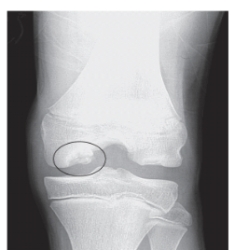
-Refer to the figure. What is the defect in the circle? Hint: This is the most common location for this lesion.
A) Osteochondritis dissecans
B) Fracture
C) Ewing's sarcoma
D) Osgood-Schlatter disease
Question
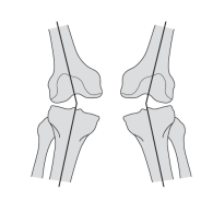
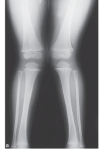
-Refer to the figure. The developmental deformity shown here is described as:
A) Bilateral genu varum
B) Bilateral genu valgus
C) Ipsilateral genu valgus
D) Contralateral genu varum

Unlock Deck
Sign up to unlock the cards in this deck!
Unlock Deck
Unlock Deck
1/10
Play
Full screen (f)
Deck 13: Radiologic Evaluation of the Knee
1
Which of following views provides the best visualization of the patella and patellofemoral joint?
A) Anteroposterior
B) Lateral
C) PA axial "tunnel"
D) Tangential view of the patellofemoral joint
A) Anteroposterior
B) Lateral
C) PA axial "tunnel"
D) Tangential view of the patellofemoral joint
Tangential view of the patellofemoral joint
2
Bony avulsion of the tibial attachment of the anterior cruciate ligament (ACL) is likely to be best visualized on which radiograph view?
A) Anteroposterior
B) Lateral
C) PA axial "tunnel"
D) Tangential view of the patellofemoral joint
A) Anteroposterior
B) Lateral
C) PA axial "tunnel"
D) Tangential view of the patellofemoral joint
Lateral
3
Evaluation for patella alta is best performed on which radiograph view?
A) Anteroposterior
B) Lateral
C) PA axial "tunnel"
D) Tangential view of the patellofemoral joint
A) Anteroposterior
B) Lateral
C) PA axial "tunnel"
D) Tangential view of the patellofemoral joint
PA axial "tunnel"
4
Which of the following is true regarding the medial and lateral tibiofemoral joint spaces in a healthy individual with normal lower extremity alignment?
A) Lateral joint space is of greater height
B) Medial joint space is of greater height
C) Medial and lateral joint spaces are equal in height
A) Lateral joint space is of greater height
B) Medial joint space is of greater height
C) Medial and lateral joint spaces are equal in height
Unlock Deck
Unlock for access to all 10 flashcards in this deck.
Unlock Deck
k this deck
5
Lateral subluxation of the patella will be best visualized with which of the following radiograph views?
A) Anteroposterior
B) Lateral
C) PA axial "tunnel"
D) Tangential view of the patellofemoral joint
A) Anteroposterior
B) Lateral
C) PA axial "tunnel"
D) Tangential view of the patellofemoral joint
Unlock Deck
Unlock for access to all 10 flashcards in this deck.
Unlock Deck
k this deck
6
Which of the following findings warrant getting radiographs after a knee injury?
A) Inability to flex the knee to 90 degree
B) Inability to flex the knee more than 110 degrees
C) Inability to fully extend the knee
D) Inability to fully flex the knee
A) Inability to flex the knee to 90 degree
B) Inability to flex the knee more than 110 degrees
C) Inability to fully extend the knee
D) Inability to fully flex the knee
Unlock Deck
Unlock for access to all 10 flashcards in this deck.
Unlock Deck
k this deck
7
Decreased radiographic joint space, sclerosis of subchondral bone, and osteophytes are the primary features of which process?
A) Rheumatoid arthritis
B) Degenerative joint disease
C) Osgood-Schlatter disease
D) Osteochondritis dissecans
A) Rheumatoid arthritis
B) Degenerative joint disease
C) Osgood-Schlatter disease
D) Osteochondritis dissecans
Unlock Deck
Unlock for access to all 10 flashcards in this deck.
Unlock Deck
k this deck
8
-Refer to the figure. Name the view.
A) Anteroposterior
B) Lateral
C) PA axial "tunnel"
D) Tangential view of the patellofemoral joint
Unlock Deck
Unlock for access to all 10 flashcards in this deck.
Unlock Deck
k this deck
9
-Refer to the figure. What is the defect in the circle? Hint: This is the most common location for this lesion.
A) Osteochondritis dissecans
B) Fracture
C) Ewing's sarcoma
D) Osgood-Schlatter disease
Unlock Deck
Unlock for access to all 10 flashcards in this deck.
Unlock Deck
k this deck
10
-Refer to the figure. The developmental deformity shown here is described as:
A) Bilateral genu varum
B) Bilateral genu valgus
C) Ipsilateral genu valgus
D) Contralateral genu varum
Unlock Deck
Unlock for access to all 10 flashcards in this deck.
Unlock Deck
k this deck
Unlock Deck
Unlock for access to all 10 flashcards in this deck.


