Deck 11: Radiologic Evaluation of the Lumbosacral Spine and Sacroiliac Joints
Full screen (f)
Question
Question
Question
Question
Question
Question
Question
Question

-Refer to the figure. Name the view.
A) Anteroposterior view of the lumbar spine
B) Anteroposterior view of the lumbosacral spine
C) Oblique view of the lumbosacral spine
D) Lateral view of the lumbosacral spine
Question
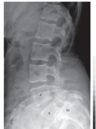
-Refer to the figure. What abnormality is present at L5-S1?
A) Spondylolisthesis
B) Spina bifida
C) Spondylosis
D) Scoliosis
Question
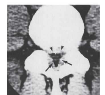
-Refer to the figure. Name the image and pathological condition.
A) Sagittal MRI, stenosis
B) Axial MRI, vertebral compression
C) Sagittal CT, spondylolisthesis
D) Axial CT, stenosis

Unlock Deck
Sign up to unlock the cards in this deck!
Unlock Deck
Unlock Deck
1/10
Play
Full screen (f)
Deck 11: Radiologic Evaluation of the Lumbosacral Spine and Sacroiliac Joints
1
Oblique radiographs of the lumbar spine are used to visualize the intervertebral foramina.
False
2
A "standard" series of lumbar radiographs would include _____________ projections.
A) anteroposterior and lateral
B) anteroposterior, lateral, and obliques
C) anteroposterior, lateral, obliques, and a lateral L5-S1
D) anteroposterior, lateral, and a lateral L5-S1
A) anteroposterior and lateral
B) anteroposterior, lateral, and obliques
C) anteroposterior, lateral, obliques, and a lateral L5-S1
D) anteroposterior, lateral, and a lateral L5-S1
anteroposterior and lateral
3
Which of the following is NOT a plane used for lumbar cross-sectional imaging?
A) Axial
B) Coronal
C) Sagittal
D) Oblique
A) Axial
B) Coronal
C) Sagittal
D) Oblique
Oblique
4
A lateral lumbar radiograph reveals a 37% anterior slippage of L5 on S1. How would this spondylolisthesis be graded?
A) Grade 1
B) Grade 2
C) Grade 3
D) Grade 4
A) Grade 1
B) Grade 2
C) Grade 3
D) Grade 4
Unlock Deck
Unlock for access to all 10 flashcards in this deck.
Unlock Deck
k this deck
5
A lumbar radiograph reveals end plate osteophytosis and vacuum disc phenomenon. What diagnosis would be representative of these findings?
A) Degenerative joint disease
B) Spondylosis
C) Degenerative disc disease
D) Diffuse idiopathic skeletal hyperostosis
A) Degenerative joint disease
B) Spondylosis
C) Degenerative disc disease
D) Diffuse idiopathic skeletal hyperostosis
Unlock Deck
Unlock for access to all 10 flashcards in this deck.
Unlock Deck
k this deck
6
If the base of a herniated disc is narrower than the apex, it is defined as:
A) Prolapsed
B) Protruded
C) Extruded
D) Herniated
A) Prolapsed
B) Protruded
C) Extruded
D) Herniated
Unlock Deck
Unlock for access to all 10 flashcards in this deck.
Unlock Deck
k this deck
7
"Bamboo spine" is the hallmark radiographic finding of this disorder.
A) Spondylosis deformans
B) Ankylosing spondylitis
C) Diffuse idiopathic skeletal hyperostosis
D) Central canal stenosis
A) Spondylosis deformans
B) Ankylosing spondylitis
C) Diffuse idiopathic skeletal hyperostosis
D) Central canal stenosis
Unlock Deck
Unlock for access to all 10 flashcards in this deck.
Unlock Deck
k this deck
8
-Refer to the figure. Name the view.
A) Anteroposterior view of the lumbar spine
B) Anteroposterior view of the lumbosacral spine
C) Oblique view of the lumbosacral spine
D) Lateral view of the lumbosacral spine
Unlock Deck
Unlock for access to all 10 flashcards in this deck.
Unlock Deck
k this deck
9
-Refer to the figure. What abnormality is present at L5-S1?
A) Spondylolisthesis
B) Spina bifida
C) Spondylosis
D) Scoliosis
Unlock Deck
Unlock for access to all 10 flashcards in this deck.
Unlock Deck
k this deck
10
-Refer to the figure. Name the image and pathological condition.
A) Sagittal MRI, stenosis
B) Axial MRI, vertebral compression
C) Sagittal CT, spondylolisthesis
D) Axial CT, stenosis
Unlock Deck
Unlock for access to all 10 flashcards in this deck.
Unlock Deck
k this deck
Unlock Deck
Unlock for access to all 10 flashcards in this deck.


