
Genetics: Analysis and Principles 5th Edition by Robert Brooker
Edition 5ISBN: 978-0073525341Genetics: Analysis and Principles 5th Edition by Robert Brooker
Edition 5ISBN: 978-0073525341 Exercise 13
When chromatin is treated with a moderate salt concentration, the linker histone H1 is removed (see Figure 10.13a). Higher salt concentration removes the rest of the histone proteins (see Figure 10.19b). If the experiment of Figure 10.12 were carried out after the DNA was treated with moderate or high salt, what would be the expected results
FIGURE 10.13 The nucleosome structure of eukaryotic chromatin as viewed by electron microscopy. The chromatin in (a) has been treated with moderate salt concentrations to remove the linker histone, H1. It exhibits the classic beads-on-a-string morphology. The chromatin in (b) has been incubated without added NaCl and shows a more compact morphology.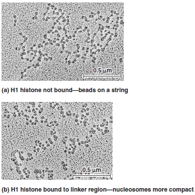
a.
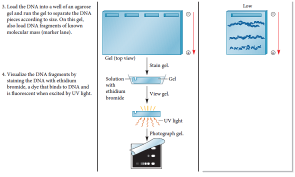
FIGURE 10.12 DNase I cuts chromatin into repeating units containing 200 bp of DNA. Starting material: Nuclei from rat liver cells.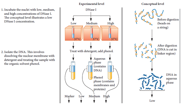
a.
FIGURE 10.19 The importance of histone proteins and scaffolding proteins in the compaction of eukaryotic chromosomes. (a) A metaphase chromosome. (b) A metaphase chromosome following treatment with high salt concentration to remove the histone proteins. The black arrow on the right points to an elongated strand of DNA. The white arrow on the left points to the scaffold (composed of nonhistone proteins), which anchors the bases of the radial loops. As shown earlier in Figure 10.18d, this scaffold contains protein filaments.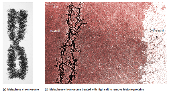
a.
FIGURE 10.18 The steps in eukaryotic chromosomal compaction leading to the metaphase chromosome.
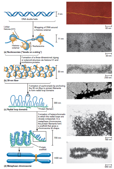
FIGURE 10.13 The nucleosome structure of eukaryotic chromatin as viewed by electron microscopy. The chromatin in (a) has been treated with moderate salt concentrations to remove the linker histone, H1. It exhibits the classic beads-on-a-string morphology. The chromatin in (b) has been incubated without added NaCl and shows a more compact morphology.
a.
FIGURE 10.12 DNase I cuts chromatin into repeating units containing 200 bp of DNA. Starting material: Nuclei from rat liver cells.
a.
FIGURE 10.19 The importance of histone proteins and scaffolding proteins in the compaction of eukaryotic chromosomes. (a) A metaphase chromosome. (b) A metaphase chromosome following treatment with high salt concentration to remove the histone proteins. The black arrow on the right points to an elongated strand of DNA. The white arrow on the left points to the scaffold (composed of nonhistone proteins), which anchors the bases of the radial loops. As shown earlier in Figure 10.18d, this scaffold contains protein filaments.
a.
FIGURE 10.18 The steps in eukaryotic chromosomal compaction leading to the metaphase chromosome.
Explanation Verified
Chromatin:
Chromatin is a combination o...
Genetics: Analysis and Principles 5th Edition by Robert Brooker
Why don’t you like this exercise?
Other Minimum 8 character and maximum 255 character
Character 255


