
Genetics: Analysis and Principles 5th Edition by Robert Brooker
Edition 5ISBN: 978-0073525341Genetics: Analysis and Principles 5th Edition by Robert Brooker
Edition 5ISBN: 978-0073525341 Exercise 19
The technique of dideoxy sequencing of DNA is described in Chapter 20. The technique relies on the use of dideoxyribonucleotides (shown in Figures 20.11 and 20.12). A dideoxyribonucleotide has a hydrogen atom attached to the 3 -carbon atom instead of an OH group. When a dideoxyribonucleotide is incorporated into a newly made strand, the strand cannot grow any longer. Explain why.FIGURE 20.11 The structure of a dideoxyribonucleotide. Note that the 3 group is a hydrogen rather than an -OH group. For this reason, another nucleotide cannot be attached at the 3 position. 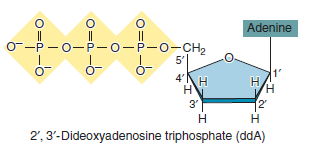
a.
FIGURE 20.12 The protocol for DNA sequencing by the dideoxy method. (a) The method begins with DNA in which the target DNA has been inserted into a vector. For the primer to bind, the recombinant vector must be denatured into single-stranded DNA at the beginning of the experiment. Only the strand needed for DNA sequencing is shown here. This diagram schematically depicts a series of bands on a gel; the four colors of the bands occur because each type of dideoxyribonucleotide is labeled with a different-colored fluorescent molecule. As each band passes a laser, the fluorescent dye is excited by the laser beam, and the fluorescence emission is recorded by a fluorescence detector. The detector reads the level of fluorescence at four wavelengths corresponding to the four dyes. (b) As shown in the printout, the peaks of fluorescence correspond to the DNA sequence that is complementary to the target DNA.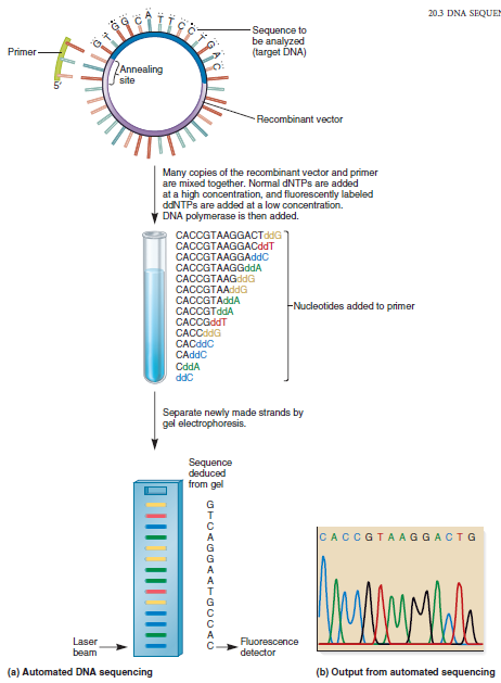
Explanation Verified
The DNA strand cannot grow any longer si...
Genetics: Analysis and Principles 5th Edition by Robert Brooker
Why don’t you like this exercise?
Other Minimum 8 character and maximum 255 character
Character 255


