
Genetics: Analysis and Principles 5th Edition by Robert Brooker
Edition 5ISBN: 978-0073525341Genetics: Analysis and Principles 5th Edition by Robert Brooker
Edition 5ISBN: 978-0073525341 Exercise 17
Describe the structure of a polysome, which is depicted in Figure 13.13a.FIGURE 13.13 Ribosomal structure. (a) Electron micrograph of ribosomes in the act of translation. Ribosomes are blue, mRNA is red, and polypeptides are green. (b) Crystal structure of the 50S and 30S subunits in bacteria. The rRNA is shown in gray strands (50S subunit) and turquoise strands (30S subunit), and proteins are shown in magenta (50S subunit) and navy blue (30S subunit). (c) A model depicting the sites where tRNA and mRNA bind to an intact ribosome. The mRNA lies on the surface of the 30S subunit. The E, P, and A sites are formed at the interface between the large and small subunits. The growing polypeptide exits through a hole in the 50S subunit. 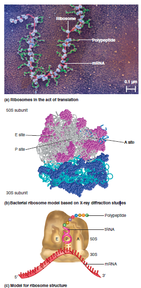
Explanation Verified
In this question, we discuss the structu...
Genetics: Analysis and Principles 5th Edition by Robert Brooker
Why don’t you like this exercise?
Other Minimum 8 character and maximum 255 character
Character 255


