
Essentials of the Living World 5th Edition by George Johnson
Edition 5ISBN: 978-0078096945Essentials of the Living World 5th Edition by George Johnson
Edition 5ISBN: 978-0078096945 Exercise 2
Are New Microtubules Made When the Spindle Forms?
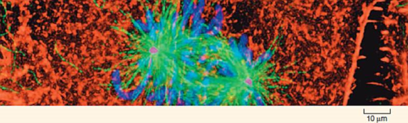
During interphase, before the beginning of meiosis, only a few long microtubules extend from the so-called centrosome (a zone around the centrioles of animal cells where microtubules are organized) to the cell periphery. Like most microtubules, they are refreshed at a low rate with resynthesis. Late in prophase, however, a dramatic change can be seen: the centrosome appears to divide into two, and a large increase is seen in the number of microtubules radiating from each of the two daughter centrosomes. The two clusters of new microtubules are easily seen as the green fibers connecting to the two sets of purple chromosomes in the micrograph of early prophase below (a micrograph is a photo taken through a microscope). This burst of microtubule assembly marks the beginning of the formation of the spindle characteristic of prophase and metaphase. When these clusters of new microtubules first became known to cell biologists, they asked whether these were existing microtubules being repositioned in the spindle or newly synthesized microtubules.
The graph to the upper right displays the results of an experiment designed to answer this question. Mammalian cells in culture (cells in culture are growing in the laboratory on artificial medium) were injected with microtubule subunits (tubulin) to which a fluorescent dye had been attached (a fluorescent dye is one that glows when exposed to ultraviolet or short-wavelength visual light). After the fluorescent subunits had become incorporated into the cells' microtubules, all the fluorescence in a small region of a cell was bleached by an intense laser beam, destroying the microtubules there. Any subsequent rebuilding of microtubules in the bleached region would have to employ the fluorescent subunits present in the cell, causing recovery of fluorescence in the bleached region. The graph reports this recovery as a function of time for interphase and metaphase cells. The dotted line represents the time for 50% recovery of fluorescence (t 1/2 ) (that is, t 1/2 is the time required for half of the microtubules in the region to be resynthesized).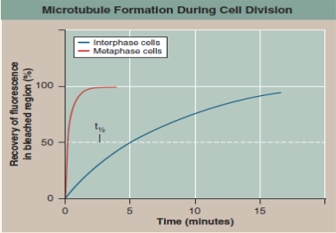
Interpreting Data Is there a difference in the rate at which microtubules are synthesized during interphase and metaphase? How big is the difference? What might account for it?
Explanation Verified
Microtubules are made of a protein calle...
Essentials of the Living World 5th Edition by George Johnson
Why don’t you like this exercise?
Other Minimum 8 character and maximum 255 character
Character 255


