Solved

Passage The K-Ras Gene Codes for a Small Guanine Nucleotide-Binding Protein

Question 155

Multiple Choice

Passage
The K-ras gene codes for a small guanine nucleotide-binding protein, which acts as a molecular activator in numerous signaling pathways that promote DNA replication along with cell growth and survival.  K-ras mutations at codons 12 and 13 function as biological markers for diagnosis of tumors.  The DNA of SW-480 cells, a line originating from a colon adenocarcinoma, is the standard positive control used to identify codon 12 mutations in K-ras testing.
Passage The K-ras gene codes for a small guanine nucleotide-binding protein, which acts as a molecular activator in numerous signaling pathways that promote DNA replication along with cell growth and survival.  K-ras mutations at codons 12 and 13 function as biological markers for diagnosis of tumors.  The DNA of SW-480 cells, a line originating from a colon adenocarcinoma, is the standard positive control used to identify codon 12 mutations in K-ras testing.    <strong>Figure 1</strong>  Structural comparison of K-ras codon 12 in wild-type cells and SW-480 cellsPCR sequence analysis has been the preferred method for studying genetic mutations such as those in K-ras.  However, challenges in analyzing PCR amplicons have prompted the development of DNA melting curve analysis (MCA) .  In the MCA technique, DNA is extracted from clinical samples and amplified by PCR.  The amplified DNA is then melted (30-70°C)  and analyzed by assessing the fluorescence (at 640 nm)  of LC Green Plus, an intercalating dye that fluoresces when bound to double-stranded DNA (Figure 2) .    <strong>Figure 2</strong>  Melting curves of wild-type (WT)  and SW-480 K-ras DNA.To study K-ras activity in colorectal cancer cells, short tandem repeats (STRs)  were introduced into the K-ras gene.  STRs are short stretches of DNA with repeating nucleotides, and four different STR sequences were inserted into the K-ras gene and analyzed via MCA under different experimental conditions (Table 1) .<strong>Table 1</strong>  Sequences Studied in K-ras DNA MCA Analysis.    Liu YP, Wu HY, Yang X, et al. Diagnostic accuracy of high resolution melting analysis for detection of KRAS mutations: a systematic review and meta-analysis. Sci Rep. 2014;4:7521. -Assuming that additional mutations similar to the one shown in Figure 1 occur throughout the mutant K-ras gene, what would be the best explanation for the difference in denaturation between wild-type (WT)  and mutant K-ras DNA? A) Mutant DNA has a higher GC content than WT DNA. B) Mutant DNA has a lower GC content than WT DNA. C) WT DNA is longer than mutant DNA. D) WT DNA is shorter than mutant DNA. 
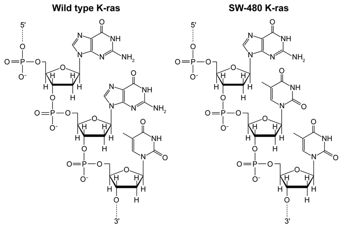
Figure 1  Structural comparison of K-ras codon 12 in wild-type cells and SW-480 cellsPCR sequence analysis has been the preferred method for studying genetic mutations such as those in K-ras.  However, challenges in analyzing PCR amplicons have prompted the development of DNA melting curve analysis (MCA) .  In the MCA technique, DNA is extracted from clinical samples and amplified by PCR.  The amplified DNA is then melted (30-70°C) and analyzed by assessing the fluorescence (at 640 nm) of LC Green Plus, an intercalating dye that fluoresces when bound to double-stranded DNA (Figure 2) .
Passage The K-ras gene codes for a small guanine nucleotide-binding protein, which acts as a molecular activator in numerous signaling pathways that promote DNA replication along with cell growth and survival.  K-ras mutations at codons 12 and 13 function as biological markers for diagnosis of tumors.  The DNA of SW-480 cells, a line originating from a colon adenocarcinoma, is the standard positive control used to identify codon 12 mutations in K-ras testing.    <strong>Figure 1</strong>  Structural comparison of K-ras codon 12 in wild-type cells and SW-480 cellsPCR sequence analysis has been the preferred method for studying genetic mutations such as those in K-ras.  However, challenges in analyzing PCR amplicons have prompted the development of DNA melting curve analysis (MCA) .  In the MCA technique, DNA is extracted from clinical samples and amplified by PCR.  The amplified DNA is then melted (30-70°C)  and analyzed by assessing the fluorescence (at 640 nm)  of LC Green Plus, an intercalating dye that fluoresces when bound to double-stranded DNA (Figure 2) .    <strong>Figure 2</strong>  Melting curves of wild-type (WT)  and SW-480 K-ras DNA.To study K-ras activity in colorectal cancer cells, short tandem repeats (STRs)  were introduced into the K-ras gene.  STRs are short stretches of DNA with repeating nucleotides, and four different STR sequences were inserted into the K-ras gene and analyzed via MCA under different experimental conditions (Table 1) .<strong>Table 1</strong>  Sequences Studied in K-ras DNA MCA Analysis.    Liu YP, Wu HY, Yang X, et al. Diagnostic accuracy of high resolution melting analysis for detection of KRAS mutations: a systematic review and meta-analysis. Sci Rep. 2014;4:7521. -Assuming that additional mutations similar to the one shown in Figure 1 occur throughout the mutant K-ras gene, what would be the best explanation for the difference in denaturation between wild-type (WT)  and mutant K-ras DNA? A) Mutant DNA has a higher GC content than WT DNA. B) Mutant DNA has a lower GC content than WT DNA. C) WT DNA is longer than mutant DNA. D) WT DNA is shorter than mutant DNA. 
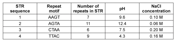
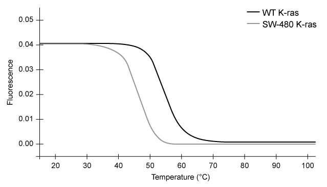
Figure 2  Melting curves of wild-type (WT) and SW-480 K-ras DNA.To study K-ras activity in colorectal cancer cells, short tandem repeats (STRs) were introduced into the K-ras gene.  STRs are short stretches of DNA with repeating nucleotides, and four different STR sequences were inserted into the K-ras gene and analyzed via MCA under different experimental conditions (Table 1) .Table 1  Sequences Studied in K-ras DNA MCA Analysis.
Passage The K-ras gene codes for a small guanine nucleotide-binding protein, which acts as a molecular activator in numerous signaling pathways that promote DNA replication along with cell growth and survival.  K-ras mutations at codons 12 and 13 function as biological markers for diagnosis of tumors.  The DNA of SW-480 cells, a line originating from a colon adenocarcinoma, is the standard positive control used to identify codon 12 mutations in K-ras testing.    <strong>Figure 1</strong>  Structural comparison of K-ras codon 12 in wild-type cells and SW-480 cellsPCR sequence analysis has been the preferred method for studying genetic mutations such as those in K-ras.  However, challenges in analyzing PCR amplicons have prompted the development of DNA melting curve analysis (MCA) .  In the MCA technique, DNA is extracted from clinical samples and amplified by PCR.  The amplified DNA is then melted (30-70°C)  and analyzed by assessing the fluorescence (at 640 nm)  of LC Green Plus, an intercalating dye that fluoresces when bound to double-stranded DNA (Figure 2) .    <strong>Figure 2</strong>  Melting curves of wild-type (WT)  and SW-480 K-ras DNA.To study K-ras activity in colorectal cancer cells, short tandem repeats (STRs)  were introduced into the K-ras gene.  STRs are short stretches of DNA with repeating nucleotides, and four different STR sequences were inserted into the K-ras gene and analyzed via MCA under different experimental conditions (Table 1) .<strong>Table 1</strong>  Sequences Studied in K-ras DNA MCA Analysis.    Liu YP, Wu HY, Yang X, et al. Diagnostic accuracy of high resolution melting analysis for detection of KRAS mutations: a systematic review and meta-analysis. Sci Rep. 2014;4:7521. -Assuming that additional mutations similar to the one shown in Figure 1 occur throughout the mutant K-ras gene, what would be the best explanation for the difference in denaturation between wild-type (WT)  and mutant K-ras DNA? A) Mutant DNA has a higher GC content than WT DNA. B) Mutant DNA has a lower GC content than WT DNA. C) WT DNA is longer than mutant DNA. D) WT DNA is shorter than mutant DNA. Liu YP, Wu HY, Yang X, et al. Diagnostic accuracy of high resolution melting analysis for detection of KRAS mutations: a systematic review and meta-analysis. Sci Rep. 2014;4:7521.
-Assuming that additional mutations similar to the one shown in Figure 1 occur throughout the mutant K-ras gene, what would be the best explanation for the difference in denaturation between wild-type (WT) and mutant K-ras DNA?


A) Mutant DNA has a higher GC content than WT DNA.
B) Mutant DNA has a lower GC content than WT DNA.
C) WT DNA is longer than mutant DNA.
D) WT DNA is shorter than mutant DNA.

Correct Answer:

verifed

Verified

Unlock this answer now
Get Access to more Verified Answers free of charge

Related Questions

Unlock this Answer For Free Now!

View this answer and more for free by performing one of the following actions

qr-code

Scan the QR code to install the App and get 2 free unlocks

upload documents

Unlock quizzes for free by uploading documents