Solved

Passage Oxidative Stress Occurs in Cells When Reactive Oxygen Species (ROS)

Question 293

Multiple Choice

Passage
Oxidative stress occurs in cells when reactive oxygen species (ROS) such as H2O2, superoxide (O2) , and hydroxyl radicals (OH) interact destructively with biological molecules.  Several studies have indicated that histidine and certain peptides that contain histidine can decrease oxidative stress.A common marker of oxidative stress in cells is leakage of the enzyme lactate dehydrogenase (LDH) into the extracellular space.  Researchers incubated cultured rat cells (PC12 cells) with H2O2 in the presence of different tripeptides, each containing one alanine, one glycine, and one histidine residue.  Every possible sequence was tested, but the researchers found that the sequence GAH was the most effective in limiting extracellular LDH activity.  After obtaining these preliminary results, the following experiments were conducted.Experiment 1PC12 cells were incubated with or without 5 mM H2O2 and with varying concentrations of GAH for 4 hours.  Cells were centrifuged, and the supernatant (ie, extracellular material) was extracted and assayed for LDH activity (Figure 1) .
Passage Oxidative stress occurs in cells when reactive oxygen species (ROS)  such as H<sub>2</sub>O<sub>2</sub>, superoxide (O<sub>2</sub><sup>−</sup>) , and hydroxyl radicals (<sup>•</sup>OH)  interact destructively with biological molecules.  Several studies have indicated that histidine and certain peptides that contain histidine can decrease oxidative stress.A common marker of oxidative stress in cells is leakage of the enzyme lactate dehydrogenase (LDH)  into the extracellular space.  Researchers incubated cultured rat cells (PC12 cells)  with H<sub>2</sub>O<sub>2</sub> in the presence of different tripeptides, each containing one alanine, one glycine, and one histidine residue.  Every possible sequence was tested, but the researchers found that the sequence GAH was the most effective in limiting extracellular LDH activity.  After obtaining these preliminary results, the following experiments were conducted.Experiment 1PC12 cells were incubated with or without 5 mM H<sub>2</sub>O<sub>2</sub> and with varying concentrations of GAH for 4 hours.  Cells were centrifuged, and the supernatant (ie, extracellular material)  was extracted and assayed for LDH activity (Figure 1) .    <strong>Figure 1</strong>  Extracellular LDH activity from PC12 cells exposed to H<sub>2</sub>O<sub>2</sub> and varying concentrations of GAH.  p-values are relative to 5 mM H<sub>2</sub>O<sub>2</sub> and 0 mg/mL GAH.Experiment 2LDH was purified and assayed for activity in the presence of 5 mM H<sub>2</sub>O<sub>2</sub> and varying concentrations of GAH (Figure 2) .    <strong>Figure 2</strong>  Activity of purified LDH with varying concentrations of GAHExperiment 3PC12 cells were incubated with or without 5 mM H<sub>2</sub>O<sub>2</sub> in the presence and absence of 1 mg/mL GAH.  Cells were then stained with the fluorescent marker TUNEL, which binds strongly to apoptotic cells.  The percentage of apoptotic cells in each condition is shown in Figure 3.    <strong>Figure 3</strong>  Percentage of apoptotic cells in the presence or absence of H<sub>2</sub>O<sub>2</sub> and GAH -The purpose of measuring purified LDH activity with various amounts of GAH was most likely: A) to demonstrate that GAH does not directly inhibit LDH. B) to determine baseline extracellular LDH activity. C) to show that GAH is an effective LDH antagonist. D) to characterize the GAH-LDH binding site. 
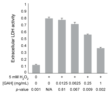
Figure 1  Extracellular LDH activity from PC12 cells exposed to H2O2 and varying concentrations of GAH.  p-values are relative to 5 mM H2O2 and 0 mg/mL GAH.Experiment 2LDH was purified and assayed for activity in the presence of 5 mM H2O2 and varying concentrations of GAH (Figure 2) .
Passage Oxidative stress occurs in cells when reactive oxygen species (ROS)  such as H<sub>2</sub>O<sub>2</sub>, superoxide (O<sub>2</sub><sup>−</sup>) , and hydroxyl radicals (<sup>•</sup>OH)  interact destructively with biological molecules.  Several studies have indicated that histidine and certain peptides that contain histidine can decrease oxidative stress.A common marker of oxidative stress in cells is leakage of the enzyme lactate dehydrogenase (LDH)  into the extracellular space.  Researchers incubated cultured rat cells (PC12 cells)  with H<sub>2</sub>O<sub>2</sub> in the presence of different tripeptides, each containing one alanine, one glycine, and one histidine residue.  Every possible sequence was tested, but the researchers found that the sequence GAH was the most effective in limiting extracellular LDH activity.  After obtaining these preliminary results, the following experiments were conducted.Experiment 1PC12 cells were incubated with or without 5 mM H<sub>2</sub>O<sub>2</sub> and with varying concentrations of GAH for 4 hours.  Cells were centrifuged, and the supernatant (ie, extracellular material)  was extracted and assayed for LDH activity (Figure 1) .    <strong>Figure 1</strong>  Extracellular LDH activity from PC12 cells exposed to H<sub>2</sub>O<sub>2</sub> and varying concentrations of GAH.  p-values are relative to 5 mM H<sub>2</sub>O<sub>2</sub> and 0 mg/mL GAH.Experiment 2LDH was purified and assayed for activity in the presence of 5 mM H<sub>2</sub>O<sub>2</sub> and varying concentrations of GAH (Figure 2) .    <strong>Figure 2</strong>  Activity of purified LDH with varying concentrations of GAHExperiment 3PC12 cells were incubated with or without 5 mM H<sub>2</sub>O<sub>2</sub> in the presence and absence of 1 mg/mL GAH.  Cells were then stained with the fluorescent marker TUNEL, which binds strongly to apoptotic cells.  The percentage of apoptotic cells in each condition is shown in Figure 3.    <strong>Figure 3</strong>  Percentage of apoptotic cells in the presence or absence of H<sub>2</sub>O<sub>2</sub> and GAH -The purpose of measuring purified LDH activity with various amounts of GAH was most likely: A) to demonstrate that GAH does not directly inhibit LDH. B) to determine baseline extracellular LDH activity. C) to show that GAH is an effective LDH antagonist. D) to characterize the GAH-LDH binding site. 
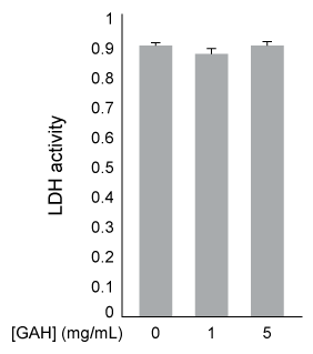
Figure 2  Activity of purified LDH with varying concentrations of GAHExperiment 3PC12 cells were incubated with or without 5 mM H2O2 in the presence and absence of 1 mg/mL GAH.  Cells were then stained with the fluorescent marker TUNEL, which binds strongly to apoptotic cells.  The percentage of apoptotic cells in each condition is shown in Figure 3.
Passage Oxidative stress occurs in cells when reactive oxygen species (ROS)  such as H<sub>2</sub>O<sub>2</sub>, superoxide (O<sub>2</sub><sup>−</sup>) , and hydroxyl radicals (<sup>•</sup>OH)  interact destructively with biological molecules.  Several studies have indicated that histidine and certain peptides that contain histidine can decrease oxidative stress.A common marker of oxidative stress in cells is leakage of the enzyme lactate dehydrogenase (LDH)  into the extracellular space.  Researchers incubated cultured rat cells (PC12 cells)  with H<sub>2</sub>O<sub>2</sub> in the presence of different tripeptides, each containing one alanine, one glycine, and one histidine residue.  Every possible sequence was tested, but the researchers found that the sequence GAH was the most effective in limiting extracellular LDH activity.  After obtaining these preliminary results, the following experiments were conducted.Experiment 1PC12 cells were incubated with or without 5 mM H<sub>2</sub>O<sub>2</sub> and with varying concentrations of GAH for 4 hours.  Cells were centrifuged, and the supernatant (ie, extracellular material)  was extracted and assayed for LDH activity (Figure 1) .    <strong>Figure 1</strong>  Extracellular LDH activity from PC12 cells exposed to H<sub>2</sub>O<sub>2</sub> and varying concentrations of GAH.  p-values are relative to 5 mM H<sub>2</sub>O<sub>2</sub> and 0 mg/mL GAH.Experiment 2LDH was purified and assayed for activity in the presence of 5 mM H<sub>2</sub>O<sub>2</sub> and varying concentrations of GAH (Figure 2) .    <strong>Figure 2</strong>  Activity of purified LDH with varying concentrations of GAHExperiment 3PC12 cells were incubated with or without 5 mM H<sub>2</sub>O<sub>2</sub> in the presence and absence of 1 mg/mL GAH.  Cells were then stained with the fluorescent marker TUNEL, which binds strongly to apoptotic cells.  The percentage of apoptotic cells in each condition is shown in Figure 3.    <strong>Figure 3</strong>  Percentage of apoptotic cells in the presence or absence of H<sub>2</sub>O<sub>2</sub> and GAH -The purpose of measuring purified LDH activity with various amounts of GAH was most likely: A) to demonstrate that GAH does not directly inhibit LDH. B) to determine baseline extracellular LDH activity. C) to show that GAH is an effective LDH antagonist. D) to characterize the GAH-LDH binding site. 
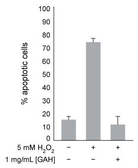
Figure 3  Percentage of apoptotic cells in the presence or absence of H2O2 and GAH
-The purpose of measuring purified LDH activity with various amounts of GAH was most likely:


A) to demonstrate that GAH does not directly inhibit LDH.
B) to determine baseline extracellular LDH activity.
C) to show that GAH is an effective LDH antagonist.
D) to characterize the GAH-LDH binding site.

Correct Answer:

verifed

Verified

Unlock this answer now
Get Access to more Verified Answers free of charge

Related Questions

Unlock this Answer For Free Now!

View this answer and more for free by performing one of the following actions

qr-code

Scan the QR code to install the App and get 2 free unlocks

upload documents

Unlock quizzes for free by uploading documents