This figure shows a portion of the mesentery and omentum removed during a work-up of a 60-year-old woman with ascites and large multicystic ovarian masses. The lesions seen here are most consistent with the diagnosis of 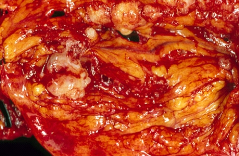
A) primary peritoneal benign tumor
B) primary peritoneal malignant tumor
C) metastatic carcinoma
D) metastatic sarcoma
E) lymphoma
Correct Answer:
Verified
Q9: Carcinoma of the esophagus most often presents
Q10: Esophageal diverticulum located above the upper esophageal
Q11: The finding of anti-endomysial antibodies in a
Q12: Which cells in the intestines are the
Q13: A 60-year-old man who complained of persistent
Q15: T-cell lymphoma was diagnosed in a 55-year-old
Q16: Which intestinal malabsorption syndrome is accompanied by
Q17: Most gastric polyps are microscopically classified as
A)
Q18: Primary tumors of the peritoneum are called
A)
Q19: Autoimmune atrophic gastritis and pernicious anemia are
Unlock this Answer For Free Now!
View this answer and more for free by performing one of the following actions

Scan the QR code to install the App and get 2 free unlocks

Unlock quizzes for free by uploading documents