This liver tumor nodule, measuring 6 cm in diameter, was seen on a computerized tomography (CT) scan of a 40-year-old man. Initially, the nodule was considered to be benign, but when it became painful, the patient decided to have it removed. Histologically, it was composed of well-differentiated hepatocytes, whereas the central scar and the fibrous septa contained proliferating bile ducts. This nodule is most likely 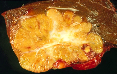
A) a hepatocellular adenoma
B) a hamartoma
C) an adenomyoma
D) a focal nodular hyperplasia
E) a hepatocellular carcinoma
Correct Answer:
Verified
Q1: Antibodies to mitochondrial dihydrolipoamide acetyltransferase are typically
Q2: Which of the following types of hepatocellular
Q3: An obese 50-year-old woman, whose height is
Q4: The most common cause of cirrhosis unrelated
Q6: Copper accumulates in the liver in Wilson
Q7: Centrolobular necrosis is typical of intoxication with
A)
Q8: These gallstones surgically removed along with the
Q9: HELLP syndrome (hemolysis, elevated liver enzymes, low
Q10: A 55-year-old man known to have drunk
Q11: Which of the following drugs causes microvesicular
Unlock this Answer For Free Now!
View this answer and more for free by performing one of the following actions

Scan the QR code to install the App and get 2 free unlocks

Unlock quizzes for free by uploading documents