Multiple Choice
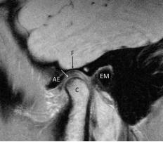
-Refer to the figure. This is a sagittal T1 MRI of a normal TMJ, made in in full occlusion. You can see the fossa (F) , articular eminence (AE) , external auditory meatus (EM) , and the condyle (C) . What is the structure the white arrow points to?
A) The superior head of the lateral pterygoid muscle
B) Articular capsule
C) Collateral ligament
D) Articular disk
Correct Answer:
Verified
Related Questions

