A 75-year-old man is brought to the emergency department due to right-sided weakness on awakening this morning. The patient has had no headache, nausea, double or blurry vision, or vertigo. Medical history includes type 2 diabetes mellitus, hypertension, and hyperlipidemia. Medications include metformin, lisinopril, and atorvastatin.
Temperature is 36.7 C (98 F) , blood pressure is 168/96 mm Hg, pulse is 92/min, and respirations are 12/min. Physical examination reveals a carotid bruit on the left. Neurologic examination shows mild dysarthria, right central facial palsy, and mild right hemiparesis.
ECG shows normal sinus rhythm. MRI of the brain reveals multiple acute cortical and subcortical infarcts in the left middle cerebral artery territory, as shown in the image below. CT angiography of the head and neck reveals 80%-90% stenosis at the origin of the left internal carotid artery. Echocardiogram shows mild left atrial enlargement, left ventricular hypertrophy, and left ventricular ejection fraction of 65%. The patient receives low-dose aspirin in the emergency department.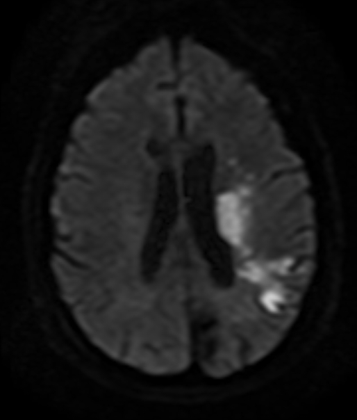
Which of the following is the most appropriate next step in management of this patient?
A) Carotid artery stenting within 2-4 months
B) Carotid endarterectomy in 1-2 weeks
C) Enoxaparin (therapeutic dose) for the duration of hospitalization
D) Long-term warfarin therapy
E) Nimodipine for 21 days
Correct Answer:
Verified
Q145: A 32-year-old right-handed woman with no significant
Q146: A 67-year-old man is brought to the
Q147: A 68-year-old right-handed man is brought by
Q148: A 57-year-old woman with known myasthenia gravis
Q149: A 73-year-old man is brought to the
Q151: A 68-year-old man is brought to the
Q152: A 38-year-old woman comes to the emergency
Q153: A 22-year-old woman is brought to the
Q154: A 32-year-old woman with a history of
Q155: A 55-year-old man comes to the emergency
Unlock this Answer For Free Now!
View this answer and more for free by performing one of the following actions

Scan the QR code to install the App and get 2 free unlocks

Unlock quizzes for free by uploading documents