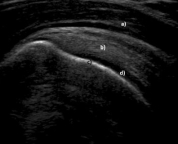
-Refer to the figure. What are the structures marked a-d in this normal longitudinal ultrasound of the shoulder?
A) (a) deltoid muscle, (b) subacromial bursa, (c) cancellous bone, (d) synovial fluid
B) (a) subacromial bursa, (b) supraspinatus muscle, (c) cortical bone, (d) articular cartilage
C) (a) subdeltoid bursa, (b) supraspinatus tendon, (c) cancellous, (d) articular capsule
D) (a) deltoid muscle, (b) supraspinatus tendon, (c) cortical bone, (d) articular cartilage
Correct Answer:
Verified
Q2: What are the differences between linear and
Q3: High signal intensity in ultrasound images is
Q4: In Doppler ultrasound, a red color indicates:
A)
Q5: Order these tissues from lowest to highest
Q6: Q7: The scanning planes in musculoskeletal ultrasound can Q8: Which is true of the imaging characteristics Q9: The advantages of ultrasound imaging include all Q10: ![]()
![]()
Unlock this Answer For Free Now!
View this answer and more for free by performing one of the following actions

Scan the QR code to install the App and get 2 free unlocks

Unlock quizzes for free by uploading documents