Figure 5.7 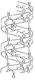
-Figure 5.7 best illustrates the
A) secondary structure of a polypeptide.
B) tertiary structure of a polypeptide.
C) quaternary structure of a protein.
D) double helix structure of DNA.
E) primary structure of a polysaccharide.
Correct Answer:
Verified
Q41: How many different kinds of polypeptides, each
Q42: The α helix and the β pleated
Q43: Which bonds are created during the formation
Q44: Refer to Figure 5.6 to answer the
Q45: What method did Frederick Sanger use to
Q47: Which type of interaction stabilizes the alpha
Q49: Refer to Figure 5.6 to answer the
Q50: Altering which of the following levels of
Q51: The R group or side chain of
Q92: ![]()
Unlock this Answer For Free Now!
View this answer and more for free by performing one of the following actions

Scan the QR code to install the App and get 2 free unlocks

Unlock quizzes for free by uploading documents