Passage
Parathyroid hormone (PTH) is an important regulator of calcium and phosphate homeostasis. Specifically, PTH functions to increase plasma calcium concentration and reduce plasma phosphate levels. PTH secretion is closely regulated by changes in plasma calcium concentration, which are detected by the human calcium-sensing receptor (hCaSR) , a G protein-coupled receptor located on chief cells of the parathyroid gland. hCaSR activation ultimately inhibits PTH release by inducing a second messenger cascade in which intracellular calcium levels rise due to the hydrolysis of the membrane phospholipid phosphatidylinositol bisphosphate (PIP2) into inositol trisphosphate (IP3) and diacylglycerol (DAG) by the membrane-bound enzyme phospholipase C (PLC) .Secondary hyperparathyroidism is a condition characterized by excessive PTH release in response to depressed serum calcium or elevated serum phosphate levels. The novel peptide AMG 416, an hCaSR agonist, is being investigated as a possible treatment for secondary hyperparathyroidism caused by end-stage renal disease (ESRD) . IP-1, a downstream metabolite of IP3, is a marker of hCaSR activity. Researchers used embryonic kidney cells lacking hCaSR (HEK-293T) and HEK-293T clone cells stably transfected with hCaSR (hCaSR) to measure IP-1 levels following AMG 416 treatment (Figure 1) .
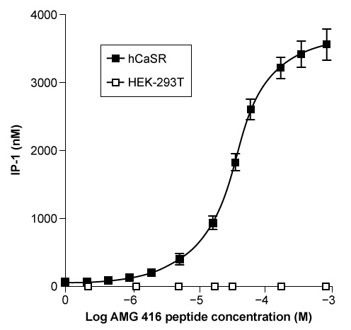 Figure 1 IP-1 concentration in embryonic kidney cells lacking hCaSR (HEK-293T) and HEK-293T clone cells transfected with hCaSR (hCaSR) following AMG 416 treatmentThe dependence of AMG 416 on extracellular calcium was investigated by incubating hCaSR cells with increasing concentrations of AMG 416 in the presence or absence of calcium treatment (Figure 2) .
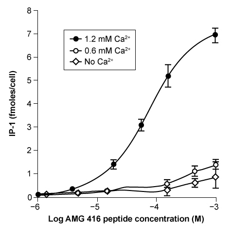 Figure 2 IP-1 levels in hCaSR cells incubated with and without calcium and AMG 416 peptide (Note: IP-1 levels are normalized to the number of cells present in each culture.) In addition, PTH concentration was measured in 5/6 Nx mice, an animal model of secondary hyperparathyroidism in human ESRD. Blood samples were taken from male and female 5/6 Nx mice for 8 hours following administration of varying concentrations of AMG 416 (Figure 3) , and PTH levels were measured.
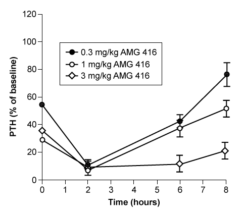 Figure 3 Serum PTH concentration following administration of AMG 416 to 5/6 Nx mice
-Assume that 5/6 Nx mice have a normal physiological response to changes in PTH secretion, and that AMG 416 treatment suppresses PTH release. Which graphic represents the blood phosphate levels that would be expected in 5/6 Nx mice eight hours after injection with indicated dosages of AMG 416?
A) 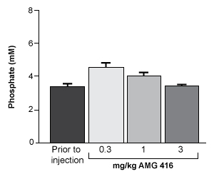
B) 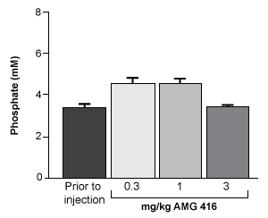
C) 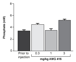
D) 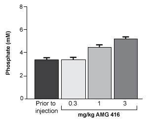
Correct Answer:
Verified
Q125: Passage
Neurulation refers to the early formation of
Q126: Passage
Osteoarthritis (OA) is a disorder marked by
Q127: Passage
Neurulation refers to the early formation of
Q128: Passage
Neurulation refers to the early formation of
Q129: Passage
Osteoarthritis (OA) is a disorder marked by
Q131: Passage
Osteoarthritis (OA) is a disorder marked by
Q132: Passage
Neurulation refers to the early formation of
Q133: Passage
Neurulation refers to the early formation of
Q134: Passage
Domestic pigs have been used as valuable
Q135: Passage
Osteoarthritis (OA) is a disorder marked by
Unlock this Answer For Free Now!
View this answer and more for free by performing one of the following actions

Scan the QR code to install the App and get 2 free unlocks

Unlock quizzes for free by uploading documents