Solved

Passage The Protozoan Euglena Gracilis Expresses Glycosyltransferases (Enzymes That Add Carbohydrates

Question 150

Multiple Choice

Passage
The protozoan Euglena gracilis expresses glycosyltransferases (enzymes that add carbohydrates to molecules) and glycosidases (enzymes that remove carbohydrates) within its membrane.  These enzymes were previously studied using radiolabeled sugars.  To avoid the use of radioactive materials, fluorescence-based assays were investigated.  Derivatives of the fluorescent molecule coumarin were attached to carbohydrates (Figure 1) for detection in fluorescence assays.Compound 1, a carbohydrate derivative with hydroxyl groups protected by benzoate (Bz) , reacted with Compound 2 to form Compound 313C NMR confirmed the formation of Compound 3 by observing a distinct C-H coupling at the anomeric carbon.  Hydroxyl deprotection and addition of Compound 4 (a coumarin analogue) gave Compound 5, a fluorescent coumarin derivative.  Compound 6, another coumarin derivative, was made following a similar sequence except an additional sugar was attached before the addition of the coumarin group.
Passage The protozoan Euglena gracilis expresses glycosyltransferases (enzymes that add carbohydrates to molecules)  and glycosidases (enzymes that remove carbohydrates)  within its membrane.  These enzymes were previously studied using radiolabeled sugars.  To avoid the use of radioactive materials, fluorescence-based assays were investigated.  Derivatives of the fluorescent molecule coumarin were attached to carbohydrates (Figure 1)  for detection in fluorescence assays.Compound <strong>1</strong>, a carbohydrate derivative with hydroxyl groups protected by benzoate (Bz) , reacted with Compound <strong>2</strong> to form Compound <strong>3</strong>.  <sup>13</sup>C NMR confirmed the formation of Compound <strong>3</strong> by observing a distinct C-H coupling at the anomeric carbon.  Hydroxyl deprotection and addition of Compound <strong>4</strong> (a coumarin analogue)  gave Compound <strong>5</strong>, a fluorescent coumarin derivative.  Compound <strong>6</strong>, another coumarin derivative, was made following a similar sequence except an additional sugar was attached before the addition of the coumarin group.    <strong>Figure 1</strong>  Synthesis of fluorescent coumarin derivatives (Note: HCT denotes the fluorescent butyl hydroxycoumarin triazole group.) Small vesicles called microsomes were isolated from Euglena gracilis membranes, and microsomal glycosyltransferase activities were investigated using a hexose donor substrate and either Compound <strong>5</strong> (Figure 2)  or Compound <strong>6</strong> (Figure 3)  as the acceptor substrate.  Thin-layer chromatography (TLC)  analysis indicated the formation of a new product in each assay.  These products were purified and characterized by liquid chromatography-mass spectrometry (LC-MS) .  The molecular ion for each product was subjected to another round of MS, in which individual hexose units (MW)  = 162)  were cleaved from the coumarin derivatives that were formed during the assay.  The MW of hydroxycoumarin triazole (HCT)  is 301.      <strong>Figure 2</strong>  Analysis of Compound <strong>5</strong> as the acceptor substrate: (A)  TLC analysis of fluorescent products; (B)  Mass spectrum of product A fragmented molecular ion peak      <strong>Figure 3</strong>  Analysis of Compound <strong>6</strong> as the acceptor substrate: (A)  TLC analysis of fluorescent products; (B)  Mass spectrum of product B fragmented molecular ion peak Adapted from: I. M. Ivanova et al.,  Fluorescent mannosides serve as acceptor substrates for glycosyltransferase and sugar-1-phosphate transferase activities in Euglena gracilis membranes.  Carbohydrate Research. © 2017 Elsevier. -  Based on the passage, which carbon gives a distinct C-H coupling in the <sup>13</sup>C NMR spectrum that confirms formation of Compound <strong>3</strong>? A) I B) II C) III D) IV 
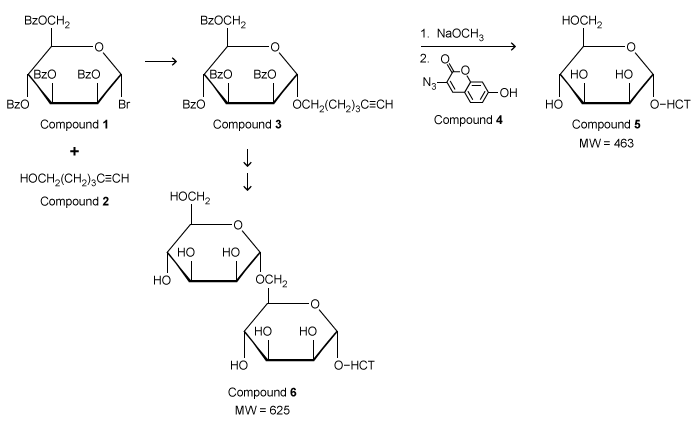
Figure 1  Synthesis of fluorescent coumarin derivatives (Note: HCT denotes the fluorescent butyl hydroxycoumarin triazole group.) Small vesicles called microsomes were isolated from Euglena gracilis membranes, and microsomal glycosyltransferase activities were investigated using a hexose donor substrate and either Compound 5 (Figure 2) or Compound 6 (Figure 3) as the acceptor substrate.  Thin-layer chromatography (TLC) analysis indicated the formation of a new product in each assay.  These products were purified and characterized by liquid chromatography-mass spectrometry (LC-MS) .  The molecular ion for each product was subjected to another round of MS, in which individual hexose units (MW) = 162) were cleaved from the coumarin derivatives that were formed during the assay.  The MW of hydroxycoumarin triazole (HCT) is 301.
Passage The protozoan Euglena gracilis expresses glycosyltransferases (enzymes that add carbohydrates to molecules)  and glycosidases (enzymes that remove carbohydrates)  within its membrane.  These enzymes were previously studied using radiolabeled sugars.  To avoid the use of radioactive materials, fluorescence-based assays were investigated.  Derivatives of the fluorescent molecule coumarin were attached to carbohydrates (Figure 1)  for detection in fluorescence assays.Compound <strong>1</strong>, a carbohydrate derivative with hydroxyl groups protected by benzoate (Bz) , reacted with Compound <strong>2</strong> to form Compound <strong>3</strong>.  <sup>13</sup>C NMR confirmed the formation of Compound <strong>3</strong> by observing a distinct C-H coupling at the anomeric carbon.  Hydroxyl deprotection and addition of Compound <strong>4</strong> (a coumarin analogue)  gave Compound <strong>5</strong>, a fluorescent coumarin derivative.  Compound <strong>6</strong>, another coumarin derivative, was made following a similar sequence except an additional sugar was attached before the addition of the coumarin group.    <strong>Figure 1</strong>  Synthesis of fluorescent coumarin derivatives (Note: HCT denotes the fluorescent butyl hydroxycoumarin triazole group.) Small vesicles called microsomes were isolated from Euglena gracilis membranes, and microsomal glycosyltransferase activities were investigated using a hexose donor substrate and either Compound <strong>5</strong> (Figure 2)  or Compound <strong>6</strong> (Figure 3)  as the acceptor substrate.  Thin-layer chromatography (TLC)  analysis indicated the formation of a new product in each assay.  These products were purified and characterized by liquid chromatography-mass spectrometry (LC-MS) .  The molecular ion for each product was subjected to another round of MS, in which individual hexose units (MW)  = 162)  were cleaved from the coumarin derivatives that were formed during the assay.  The MW of hydroxycoumarin triazole (HCT)  is 301.      <strong>Figure 2</strong>  Analysis of Compound <strong>5</strong> as the acceptor substrate: (A)  TLC analysis of fluorescent products; (B)  Mass spectrum of product A fragmented molecular ion peak      <strong>Figure 3</strong>  Analysis of Compound <strong>6</strong> as the acceptor substrate: (A)  TLC analysis of fluorescent products; (B)  Mass spectrum of product B fragmented molecular ion peak Adapted from: I. M. Ivanova et al.,  Fluorescent mannosides serve as acceptor substrates for glycosyltransferase and sugar-1-phosphate transferase activities in Euglena gracilis membranes.  Carbohydrate Research. © 2017 Elsevier. -  Based on the passage, which carbon gives a distinct C-H coupling in the <sup>13</sup>C NMR spectrum that confirms formation of Compound <strong>3</strong>? A) I B) II C) III D) IV Passage The protozoan Euglena gracilis expresses glycosyltransferases (enzymes that add carbohydrates to molecules)  and glycosidases (enzymes that remove carbohydrates)  within its membrane.  These enzymes were previously studied using radiolabeled sugars.  To avoid the use of radioactive materials, fluorescence-based assays were investigated.  Derivatives of the fluorescent molecule coumarin were attached to carbohydrates (Figure 1)  for detection in fluorescence assays.Compound <strong>1</strong>, a carbohydrate derivative with hydroxyl groups protected by benzoate (Bz) , reacted with Compound <strong>2</strong> to form Compound <strong>3</strong>.  <sup>13</sup>C NMR confirmed the formation of Compound <strong>3</strong> by observing a distinct C-H coupling at the anomeric carbon.  Hydroxyl deprotection and addition of Compound <strong>4</strong> (a coumarin analogue)  gave Compound <strong>5</strong>, a fluorescent coumarin derivative.  Compound <strong>6</strong>, another coumarin derivative, was made following a similar sequence except an additional sugar was attached before the addition of the coumarin group.    <strong>Figure 1</strong>  Synthesis of fluorescent coumarin derivatives (Note: HCT denotes the fluorescent butyl hydroxycoumarin triazole group.) Small vesicles called microsomes were isolated from Euglena gracilis membranes, and microsomal glycosyltransferase activities were investigated using a hexose donor substrate and either Compound <strong>5</strong> (Figure 2)  or Compound <strong>6</strong> (Figure 3)  as the acceptor substrate.  Thin-layer chromatography (TLC)  analysis indicated the formation of a new product in each assay.  These products were purified and characterized by liquid chromatography-mass spectrometry (LC-MS) .  The molecular ion for each product was subjected to another round of MS, in which individual hexose units (MW)  = 162)  were cleaved from the coumarin derivatives that were formed during the assay.  The MW of hydroxycoumarin triazole (HCT)  is 301.      <strong>Figure 2</strong>  Analysis of Compound <strong>5</strong> as the acceptor substrate: (A)  TLC analysis of fluorescent products; (B)  Mass spectrum of product A fragmented molecular ion peak      <strong>Figure 3</strong>  Analysis of Compound <strong>6</strong> as the acceptor substrate: (A)  TLC analysis of fluorescent products; (B)  Mass spectrum of product B fragmented molecular ion peak Adapted from: I. M. Ivanova et al.,  Fluorescent mannosides serve as acceptor substrates for glycosyltransferase and sugar-1-phosphate transferase activities in Euglena gracilis membranes.  Carbohydrate Research. © 2017 Elsevier. -  Based on the passage, which carbon gives a distinct C-H coupling in the <sup>13</sup>C NMR spectrum that confirms formation of Compound <strong>3</strong>? A) I B) II C) III D) IV 
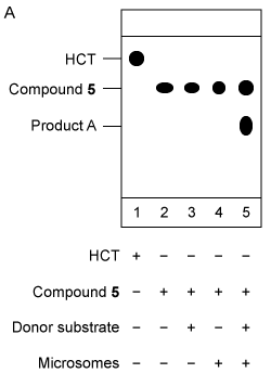
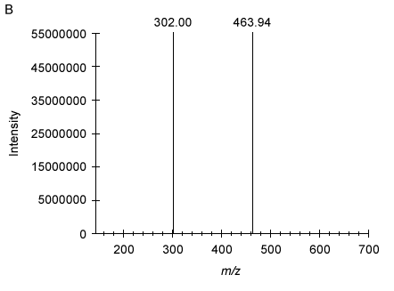
Figure 2  Analysis of Compound 5 as the acceptor substrate: (A) TLC analysis of fluorescent products; (B) Mass spectrum of product A fragmented molecular ion peak
Passage The protozoan Euglena gracilis expresses glycosyltransferases (enzymes that add carbohydrates to molecules)  and glycosidases (enzymes that remove carbohydrates)  within its membrane.  These enzymes were previously studied using radiolabeled sugars.  To avoid the use of radioactive materials, fluorescence-based assays were investigated.  Derivatives of the fluorescent molecule coumarin were attached to carbohydrates (Figure 1)  for detection in fluorescence assays.Compound <strong>1</strong>, a carbohydrate derivative with hydroxyl groups protected by benzoate (Bz) , reacted with Compound <strong>2</strong> to form Compound <strong>3</strong>.  <sup>13</sup>C NMR confirmed the formation of Compound <strong>3</strong> by observing a distinct C-H coupling at the anomeric carbon.  Hydroxyl deprotection and addition of Compound <strong>4</strong> (a coumarin analogue)  gave Compound <strong>5</strong>, a fluorescent coumarin derivative.  Compound <strong>6</strong>, another coumarin derivative, was made following a similar sequence except an additional sugar was attached before the addition of the coumarin group.    <strong>Figure 1</strong>  Synthesis of fluorescent coumarin derivatives (Note: HCT denotes the fluorescent butyl hydroxycoumarin triazole group.) Small vesicles called microsomes were isolated from Euglena gracilis membranes, and microsomal glycosyltransferase activities were investigated using a hexose donor substrate and either Compound <strong>5</strong> (Figure 2)  or Compound <strong>6</strong> (Figure 3)  as the acceptor substrate.  Thin-layer chromatography (TLC)  analysis indicated the formation of a new product in each assay.  These products were purified and characterized by liquid chromatography-mass spectrometry (LC-MS) .  The molecular ion for each product was subjected to another round of MS, in which individual hexose units (MW)  = 162)  were cleaved from the coumarin derivatives that were formed during the assay.  The MW of hydroxycoumarin triazole (HCT)  is 301.      <strong>Figure 2</strong>  Analysis of Compound <strong>5</strong> as the acceptor substrate: (A)  TLC analysis of fluorescent products; (B)  Mass spectrum of product A fragmented molecular ion peak      <strong>Figure 3</strong>  Analysis of Compound <strong>6</strong> as the acceptor substrate: (A)  TLC analysis of fluorescent products; (B)  Mass spectrum of product B fragmented molecular ion peak Adapted from: I. M. Ivanova et al.,  Fluorescent mannosides serve as acceptor substrates for glycosyltransferase and sugar-1-phosphate transferase activities in Euglena gracilis membranes.  Carbohydrate Research. © 2017 Elsevier. -  Based on the passage, which carbon gives a distinct C-H coupling in the <sup>13</sup>C NMR spectrum that confirms formation of Compound <strong>3</strong>? A) I B) II C) III D) IV Passage The protozoan Euglena gracilis expresses glycosyltransferases (enzymes that add carbohydrates to molecules)  and glycosidases (enzymes that remove carbohydrates)  within its membrane.  These enzymes were previously studied using radiolabeled sugars.  To avoid the use of radioactive materials, fluorescence-based assays were investigated.  Derivatives of the fluorescent molecule coumarin were attached to carbohydrates (Figure 1)  for detection in fluorescence assays.Compound <strong>1</strong>, a carbohydrate derivative with hydroxyl groups protected by benzoate (Bz) , reacted with Compound <strong>2</strong> to form Compound <strong>3</strong>.  <sup>13</sup>C NMR confirmed the formation of Compound <strong>3</strong> by observing a distinct C-H coupling at the anomeric carbon.  Hydroxyl deprotection and addition of Compound <strong>4</strong> (a coumarin analogue)  gave Compound <strong>5</strong>, a fluorescent coumarin derivative.  Compound <strong>6</strong>, another coumarin derivative, was made following a similar sequence except an additional sugar was attached before the addition of the coumarin group.    <strong>Figure 1</strong>  Synthesis of fluorescent coumarin derivatives (Note: HCT denotes the fluorescent butyl hydroxycoumarin triazole group.) Small vesicles called microsomes were isolated from Euglena gracilis membranes, and microsomal glycosyltransferase activities were investigated using a hexose donor substrate and either Compound <strong>5</strong> (Figure 2)  or Compound <strong>6</strong> (Figure 3)  as the acceptor substrate.  Thin-layer chromatography (TLC)  analysis indicated the formation of a new product in each assay.  These products were purified and characterized by liquid chromatography-mass spectrometry (LC-MS) .  The molecular ion for each product was subjected to another round of MS, in which individual hexose units (MW)  = 162)  were cleaved from the coumarin derivatives that were formed during the assay.  The MW of hydroxycoumarin triazole (HCT)  is 301.      <strong>Figure 2</strong>  Analysis of Compound <strong>5</strong> as the acceptor substrate: (A)  TLC analysis of fluorescent products; (B)  Mass spectrum of product A fragmented molecular ion peak      <strong>Figure 3</strong>  Analysis of Compound <strong>6</strong> as the acceptor substrate: (A)  TLC analysis of fluorescent products; (B)  Mass spectrum of product B fragmented molecular ion peak Adapted from: I. M. Ivanova et al.,  Fluorescent mannosides serve as acceptor substrates for glycosyltransferase and sugar-1-phosphate transferase activities in Euglena gracilis membranes.  Carbohydrate Research. © 2017 Elsevier. -  Based on the passage, which carbon gives a distinct C-H coupling in the <sup>13</sup>C NMR spectrum that confirms formation of Compound <strong>3</strong>? A) I B) II C) III D) IV 
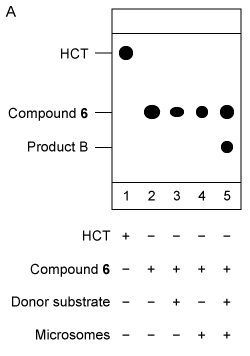
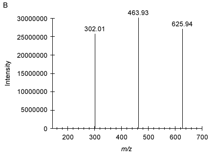
Figure 3  Analysis of Compound 6 as the acceptor substrate: (A) TLC analysis of fluorescent products; (B) Mass spectrum of product B fragmented molecular ion peak
Adapted from: I. M. Ivanova et al., "Fluorescent mannosides serve as acceptor substrates for glycosyltransferase and sugar-1-phosphate transferase activities in Euglena gracilis membranes." Carbohydrate Research. © 2017 Elsevier.
-Passage The protozoan Euglena gracilis expresses glycosyltransferases (enzymes that add carbohydrates to molecules)  and glycosidases (enzymes that remove carbohydrates)  within its membrane.  These enzymes were previously studied using radiolabeled sugars.  To avoid the use of radioactive materials, fluorescence-based assays were investigated.  Derivatives of the fluorescent molecule coumarin were attached to carbohydrates (Figure 1)  for detection in fluorescence assays.Compound <strong>1</strong>, a carbohydrate derivative with hydroxyl groups protected by benzoate (Bz) , reacted with Compound <strong>2</strong> to form Compound <strong>3</strong>.  <sup>13</sup>C NMR confirmed the formation of Compound <strong>3</strong> by observing a distinct C-H coupling at the anomeric carbon.  Hydroxyl deprotection and addition of Compound <strong>4</strong> (a coumarin analogue)  gave Compound <strong>5</strong>, a fluorescent coumarin derivative.  Compound <strong>6</strong>, another coumarin derivative, was made following a similar sequence except an additional sugar was attached before the addition of the coumarin group.    <strong>Figure 1</strong>  Synthesis of fluorescent coumarin derivatives (Note: HCT denotes the fluorescent butyl hydroxycoumarin triazole group.) Small vesicles called microsomes were isolated from Euglena gracilis membranes, and microsomal glycosyltransferase activities were investigated using a hexose donor substrate and either Compound <strong>5</strong> (Figure 2)  or Compound <strong>6</strong> (Figure 3)  as the acceptor substrate.  Thin-layer chromatography (TLC)  analysis indicated the formation of a new product in each assay.  These products were purified and characterized by liquid chromatography-mass spectrometry (LC-MS) .  The molecular ion for each product was subjected to another round of MS, in which individual hexose units (MW)  = 162)  were cleaved from the coumarin derivatives that were formed during the assay.  The MW of hydroxycoumarin triazole (HCT)  is 301.      <strong>Figure 2</strong>  Analysis of Compound <strong>5</strong> as the acceptor substrate: (A)  TLC analysis of fluorescent products; (B)  Mass spectrum of product A fragmented molecular ion peak      <strong>Figure 3</strong>  Analysis of Compound <strong>6</strong> as the acceptor substrate: (A)  TLC analysis of fluorescent products; (B)  Mass spectrum of product B fragmented molecular ion peak Adapted from: I. M. Ivanova et al.,  Fluorescent mannosides serve as acceptor substrates for glycosyltransferase and sugar-1-phosphate transferase activities in Euglena gracilis membranes.  Carbohydrate Research. © 2017 Elsevier. -  Based on the passage, which carbon gives a distinct C-H coupling in the <sup>13</sup>C NMR spectrum that confirms formation of Compound <strong>3</strong>? A) I B) II C) III D) IV 
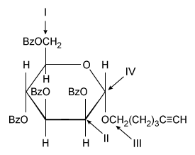
Based on the passage, which carbon gives a distinct C-H coupling in the 13C NMR spectrum that confirms formation of Compound 3?


A) I
B) II
C) III
D) IV

Correct Answer:

verifed

Verified

Unlock this answer now
Get Access to more Verified Answers free of charge

Related Questions

Unlock this Answer For Free Now!

View this answer and more for free by performing one of the following actions

qr-code

Scan the QR code to install the App and get 2 free unlocks

upload documents

Unlock quizzes for free by uploading documents